Download Mendeley
Development of a drug delivery vehicle protein exhibiting high binding affinity and low leakage of the anti-cancer drug SN-38.
Nakatsuji, M., Muroya, H., Okubo, R., Teraoka, Y., Yamada, M., Nishide, K., Yoshida, H., Furuta, K., Koyama, R., Kida, T., Doi, H., Nishimura, S., Inui, T.(2026) Int J Biol Macromol 381: 154313-154313
- PubMed: 42680028 Search on PubMed
- DOI: https://doi.org/10.1016/j.ijbiomac.2026.154313
- Primary Citation Related Structures:
26JH, 26RW - PubMed Abstract:
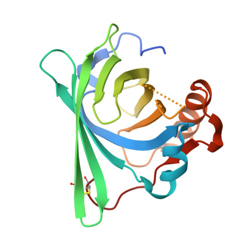
Drug leakage from delivery vehicles is a major limitation of drug delivery systems (DDSs) for cancer chemotherapy because premature release of loaded drugs reduces therapeutic efficacy and increases off-target toxicity. We previously developed a DDS for the poorly water-soluble anti-cancer drug SN-38 using lipocalin-type prostaglandin D synthase (L-PGDS). To suppress drug leakage, in this study we generated an L-PGDS mutant (M94W-M145W) with enhanced binding affinity for SN-38 by introducing amino acid substitutions into the ligand-binding cavity. Docking simulations identified residues involved in SN-38 recognition, and selected residues were replaced with tryptophan to strengthen ligand binding. The dissociation constant of the M94W-M145W mutant for SN-38 was 2.7 ± 0.4 μM, approximately 4-fold lower than that of L-PGDS. In addition, 1 mM M94W-M145W enhanced the solubility of SN-38 by approximately 3.3-fold compared with 1 mM L-PGDS. In vitro release assays showed that the SN-38/M94W-M145W complex released SN-38 more slowly than the SN-38/L-PGDS complex. We also determined the crystal structure of the 10-O-(3-fluoropropyl)-substituted SN-38 derivative/M94W-M145W complex. The overall structure of M94W-M145W retained the typical lipocalin fold, indicating that these substitutions do not alter the global protein architecture. Two SN-38 derivative molecules were accommodated within the cavity through hydrogen bonding and hydrophobic interactions, including π-π stacking interactions introduced by the substituted tryptophan residues. These findings demonstrate that simple amino acid substitutions in L-PGDS can optimize drug binding, improve solubility, and suppress drug release, thus providing a basis for affinity-driven design of protein-based DDSs.
- Department of Applied Life Sciences, Graduate School of Life and Environmental Sciences, Osaka Prefecture University, 1-1 Gakuen-cho, Naka-ku, Sakai, Osaka, 599-8531, Japan; Department of Pathobiochemistry, Faculty of Pharmacy, Osaka Medical and Pharmaceutical University, 4-20-1 Nasahara, Takatsuki, Osaka, 569-1094, Japan.
Organizational Affiliation: