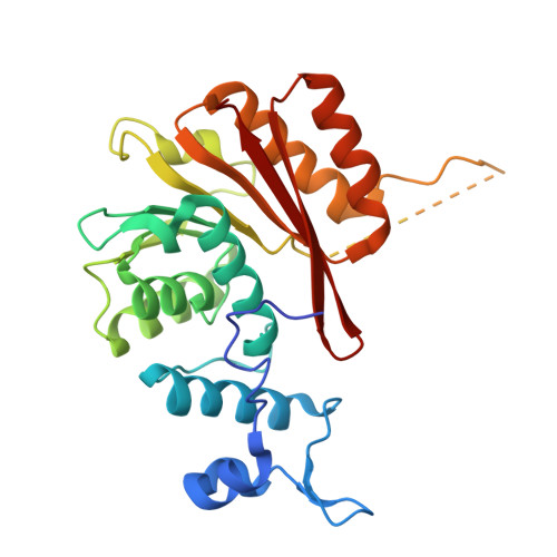
28ZC | pdb_000028zc

Crystal structure of the human METTL16 Methyltransferase Domain in complex with a bisubstrate analogue (BA4)


Experimental Data Snapshot

  • Method: X-RAY DIFFRACTION
  • Resolution: 2.17 Å
  • R-Value Free: 
    0.234 (Depositor), 0.235 (DCC) 
  • R-Value Work: 
    0.207 (Depositor), 0.208 (DCC) 
  • R-Value Observed: 
    0.209 (Depositor) 

Starting Model: experimental
View more details

wwPDB Validation 3D Report Full Report

Validation slider image for 28ZC

Ligand Structure Quality Assessment 


This is version 1.2 of the entry. See complete history. 

Literature

Catalysis tuned for metabolic sensing: dynamics and mechanism of the RNA methyltransferase METTL16.

Stamm, F., Wan, Z., Vargas-Rosales, P.A., Corbeski, I., Braud, E., Iannazzo, L., Cui, Q., Caflisch, A.

(2026) RSC Chem Biol 

  • DOI: https://doi.org/10.1039/d6cb00242k
  • Primary Citation Related Structures: 
    28WE, 28ZC

  • PubMed Abstract: 

    The methyltransferase-like protein 16 (METTL16) is the writer enzyme that methylates adenosine to form N6-methyladenosine (m6A) on U6 spliceosomal RNA and MAT2A mRNA. The latter encodes the enzyme responsible for the synthesis of S -adenosyl methionine (SAM). Since methylation influences the stability of MAT2A mRNA, METTL16 is the sensor of SAM in the cell. Here, we investigate the (co)substrate recognition and catalytic mechanism of METTL16 by a combination of in vitro and simulation methods. We first solved the crystal structures of METTL16 in complex with bisubstrate analogs (BAs), conjugates of a SAM-like moiety connected to the N6-atom of adenosine. The METTL16/BA crystal structures provide an atomistic picture of a transition-state mimic of methyl transfer. Molecular dynamics (MD) simulations started from complexes with oligonucleotide-RNA modeled on the METTL16/BAs structures show rapid unbinding of SAM. The presence of the MAT2A hairpin 1 RNA stabilizes SAM in its recognition pocket, and substantially reduces the intrinsic flexibility of the R-loop and N-terminal segment of METTL16. Quantum mechanics/molecular mechanics (QM/MM) free energy calculations provide evidence that methyl transfer occurs without prior deprotonation of adenosine-N6. Furthermore, the simulations indicate that electrostatic interactions between the charged groups of SAM and the catalytic pocket are less pronounced for METTL16 than for METTL3-14, which is the main m6A-RNA writer enzyme. The multidisciplinary approach used here sheds light on the intrinsic plasticity of METTL16, the rigidification upon RNA-substrate binding, the catalytic mechanism, and (co)product release. Overall, our study suggests that the latter step is rate-limiting.


  • Organizational Affiliation: 
    • Department of Biochemistry, University of Zurich Winterthurerstrasse 190 8057 Zürich Switzerland caflisch@bioc.uzh.ch.

Macromolecule Content 

  • Total Structure Weight: 34.77 kDa 
  • Atom Count: 2,151 
  • Modeled Residue Count: 259 
  • Deposited Residue Count: 292 
  • Unique protein chains: 1

Macromolecules

Find similar proteins by:|  3D Structure
Entity ID: 1
MoleculeChains  Sequence LengthOrganismDetailsImage
RNA N(6)-adenosine-methyltransferase METTL16292Homo sapiensMutation(s): 0 
Gene Names: METTL16, METT10D
EC: 2.1.1.348 (PDB Primary Data), 2.1.1.346 (PDB Primary Data)
UniProt & NIH Common Fund Data Resources
Find proteins for Q86W50 (Homo sapiens)
Explore Q86W50 
Go to UniProtKB:  Q86W50
PHAROS:  Q86W50
GTEx:  ENSG00000127804 
Entity Groups
Sequence Clusters30% Identity50% Identity70% Identity90% Identity95% Identity100% Identity
UniProt GroupQ86W50
Sequence Annotations
Expand
Reference Sequence

Small Molecules

Ligands 4 Unique
IDChains Name / Formula / InChI Key2D Diagram3D Interactions
I0C
(Subject of Investigation/LOI)

Query on I0C


Download:Ideal Coordinates CCD File
I [auth A](2~{S})-4-[[(2~{R},3~{S},4~{R},5~{R})-5-(6-aminopurin-9-yl)-3,4-bis(oxidanyl)oxolan-2-yl]methyl-[3-[[9-[(2~{R},5~{R})-5-(hydroxymethyl)-3,4-bis(oxidanyl)oxolan-2-yl]-7~{H}-purin-6-yl]amino]propyl]amino]-2-azanyl-butanoic acid
C27 H38 N12 O9
BNMNWGQODJWABC-MAYUJHHQSA-N
PO4

Query on PO4


Download:Ideal Coordinates CCD File
J [auth A],
K [auth A]
PHOSPHATE ION
O4 P
NBIIXXVUZAFLBC-UHFFFAOYSA-K
EDO

Query on EDO


Download:Ideal Coordinates CCD File
B [auth A]
C [auth A]
D [auth A]
E [auth A]
F [auth A]
B [auth A],
C [auth A],
D [auth A],
E [auth A],
F [auth A],
G [auth A],
H [auth A]
1,2-ETHANEDIOL
C2 H6 O2
LYCAIKOWRPUZTN-UHFFFAOYSA-N
CL

Query on CL


Download:Ideal Coordinates CCD File
L [auth A],
M [auth A]
CHLORIDE ION
Cl
VEXZGXHMUGYJMC-UHFFFAOYSA-M

Experimental Data & Validation

Experimental Data

  • Method: X-RAY DIFFRACTION
  • Resolution: 2.17 Å
  • R-Value Free:  0.234 (Depositor), 0.235 (DCC) 
  • R-Value Work:  0.207 (Depositor), 0.208 (DCC) 
  • R-Value Observed: 0.209 (Depositor) 
Space Group: I 41 3 2
Unit Cell:
Length ( Å )Angle ( ˚ )
a = 188.783α = 90
b = 188.783β = 90
c = 188.783γ = 90
Software Package:
Software NamePurpose
PHENIXrefinement
autoPROCdata scaling
PHASERphasing
PDB_EXTRACTdata extraction
autoPROCdata reduction

Structure Validation

View Full Validation Report


Ligand Structure Quality Assessment 


Entry History 

& Funding Information

Deposition Data


Funding OrganizationLocationGrant Number
Swiss National Science FoundationSwitzerland--

Revision History  (Full details and data files)

  • Version 1.0: 2026-10-14
    Type: Initial release
  • Version 1.1: 2026-10-21
    Changes: Database references
  • Version 1.2: 2026-11-04
    Changes: Database references