30KD | pdb_000030kd

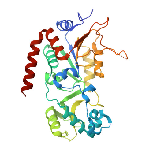
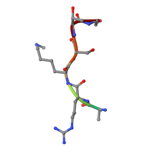
Crystal structure of human SIRT2 in complex with KMyrMe peptide


Experimental Data Snapshot

  • Method: X-RAY DIFFRACTION
  • Resolution: 1.45 Å
  • R-Value Free: 
    0.199 (Depositor), 0.199 (DCC) 
  • R-Value Work: 
    0.174 (Depositor), 0.174 (DCC) 
  • R-Value Observed: 
    0.175 (Depositor) 

Starting Model: experimental
View more details

wwPDB Validation 3D Report Full Report

Validation slider image for 30KD

Ligand Structure Quality Assessment 


This is version 1.1 of the entry. See complete history. 

Literature

Structural and Enzymatic Basis for Impaired Deacylation of N-Acyl-N-Methyl Lysine Residues.

Hilscher, S., Bottcher, P.H., Friedrich, F., Pannek, M., Baselious, F., Meleshin, M., Einsle, O., Barinka, C., Steegborn, C., Sippl, W., Jung, M., Schutkowski, M., Schiene-Fischer, C.

(2026) Biochemistry 65: 3092-3105

  • DOI: https://doi.org/10.1021/acs.biochem.6c00508
  • Primary Citation Related Structures: 
    30KD, 30KE

  • PubMed Abstract: 

    Posttranslational lysine acetylation and methylation are generally considered mutually exclusive modifications of the ε-amino group. The recent discovery of N-acetyl-N-methyl lysine (KAcMe) on histone H4 showed that both marks can coexist on the same lysine side chain, raising the question of whether this dual modification is enzymatically reversible. Here, we synthesized model peptides containing N-acyl lysine or N-acyl-N-methyl lysine residues and tested their processing by Zn2+-dependent HDACs and NAD+-dependent sirtuins. While the corresponding unmethylated substrates were deacylated by the expected enzymes, KAcMe-containing peptides were not detectably deacetylated by either enzyme family. N-methylation also blocked HDAC8/11-mediated demyristoylation and SIRT5-mediated desuccinylation, whereas SIRT2 and SIRT6 retained weak demyristoylase activity toward N-myristoyl-N-methyl lysine. X-ray crystal structures of sirtuin-peptide complexes, supported by inhibition studies and molecular docking, reveal that N-methylation disrupts productive substrate positioning and misaligns the scissile amide bond for catalysis. These findings suggest that KAcMe is resistant to canonical lysine deacetylases and may represent a persistent chromatin-associated modification.


  • Organizational Affiliation: 
    • Department of Medicinal Chemistry, Institute of Pharmacy, Martin-Luther-University Halle-Wittenberg, Kurt-Mothes-Str. 3, Halle/Saale06120, Germany.

Macromolecule Content 

  • Total Structure Weight: 35.65 kDa 
  • Atom Count: 2,745 
  • Modeled Residue Count: 296 
  • Deposited Residue Count: 311 
  • Unique protein chains: 2

Macromolecules

Find similar proteins by:|  3D Structure
Entity ID: 1
MoleculeChains  Sequence LengthOrganismDetailsImage
NAD-dependent protein deacetylase sirtuin-2304Homo sapiensMutation(s): 0 
Gene Names: SIRT2, SIR2L, SIR2L2
EC: 3.5.1 (PDB Primary Data), 2.3.1 (UniProt), 2.3.1.286 (UniProt)
UniProt & NIH Common Fund Data Resources
Find proteins for Q8IXJ6 (Homo sapiens)
Explore Q8IXJ6 
Go to UniProtKB:  Q8IXJ6
GTEx:  ENSG00000068903 
Entity Groups
Sequence Clusters30% Identity50% Identity70% Identity90% Identity95% Identity100% Identity
UniProt GroupQ8IXJ6
Sequence Annotations
Expand
Reference Sequence
Find similar proteins by:  Sequence   |   3D Structure  
Entity ID: 2
MoleculeChains  Sequence LengthOrganismDetailsImage
KMyrMe peptideB [auth C]7synthetic constructMutation(s): 0 
Sequence Annotations
Expand
Reference Sequence

Small Molecules

Modified Residues  1 Unique
IDChains Modified ResidueModification TypeFormula2D Diagram
MLZ
Query on MLZ
LYSMethylationC7 H16 N2 O2

Experimental Data & Validation

Experimental Data

  • Method: X-RAY DIFFRACTION
  • Resolution: 1.45 Å
  • R-Value Free:  0.199 (Depositor), 0.199 (DCC) 
  • R-Value Work:  0.174 (Depositor), 0.174 (DCC) 
  • R-Value Observed: 0.175 (Depositor) 
Space Group: P 1 21 1
Unit Cell:
Length ( Å )Angle ( ˚ )
a = 36.108α = 90
b = 73.117β = 95.49
c = 55.017γ = 90
Software Package:
Software NamePurpose
PHENIXrefinement
Aimlessdata scaling
PDB_EXTRACTdata extraction
autoPROCdata reduction
PHASERphasing

Structure Validation

View Full Validation Report


Ligand Structure Quality Assessment 


Entry History 

& Funding Information

Deposition Data


Funding OrganizationLocationGrant Number
German Research Foundation (DFG)GermanyJu295/18-1

Revision History  (Full details and data files)

  • Version 1.0: 2026-09-30
    Type: Initial release
  • Version 1.1: 2026-11-04
    Changes: Database references