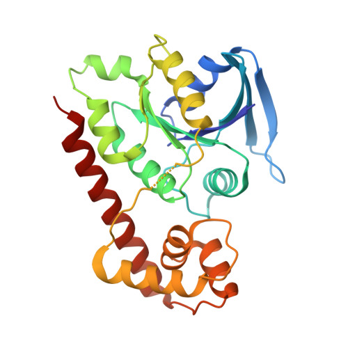
3A1U | pdb_00003a1u

Crystal structue of the cytosolic domain of T. maritima FeoB iron iransporter in GMPPNP form


Experimental Data Snapshot

  • Method: X-RAY DIFFRACTION
  • Resolution: 1.80 Å
  • R-Value Free: 
    0.219 (Depositor), 0.220 (DCC) 
  • R-Value Work: 
    0.183 (Depositor), 0.180 (DCC) 
  • R-Value Observed: 
    0.185 (Depositor) 

Starting Model: experimental
View more details

wwPDB Validation   3D Report Full Report


Ligand Structure Quality Assessment 


This is version 1.2 of the entry. See complete history


Literature

Structural basis of novel interactions between the small-GTPase and GDI-like domains in prokaryotic FeoB iron transporter

Hattori, M.Jin, Y.Nishimasu, H.Tanaka, Y.Mochizuki, M.Uchiumi, T.Ishitani, R.Ito, K.Nureki, O.

(2009) Structure 17: 1345-1355

  • DOI: https://doi.org/10.1016/j.str.2009.08.007
  • Primary Citation of Related Structures:  
    3A1S, 3A1T, 3A1U, 3A1V, 3A1W

  • PubMed Abstract: 

    The FeoB family proteins are widely distributed prokaryotic membrane proteins involved in Fe(2+) uptake. FeoB consists of N-terminal cytosolic and C-terminal transmembrane domains. The N-terminal region of the cytosolic domain is homologous to small GTPase (G) proteins and is considered to regulate Fe(2+) uptake. The spacer region connecting the G and TM domains reportedly functions as a GDP dissociation inhibitor (GDI)-like domain that stabilizes the GDP-binding state. However, the function of the G and GDI-like domains in iron uptake remains unclear. Here, we report the structural and functional analyses of the FeoB cytosolic domain from Thermotoga maritima. The structure-based mutational analysis indicated that the interaction between the G and GDI-like domains is important for both the GDI and Fe(2+) uptake activities. On the basis of these results, we propose a regulatory mechanism of Fe(2+) uptake.


  • Organizational Affiliation
    • Department of Basic Medical Sciences, Institute of Medical Science, The University of Tokyo, Minato-ku, Tokyo, Japan.

Macromolecules
Find similar proteins by:  (by identity cutoff)  |  3D Structure
Entity ID: 1
MoleculeChains Sequence LengthOrganismDetailsImage
Iron(II) transport protein B
A, B
258Thermotoga maritimaMutation(s): 0 
Gene Names: TM0051
UniProt
Find proteins for Q9WXQ8 (Thermotoga maritima (strain ATCC 43589 / DSM 3109 / JCM 10099 / NBRC 100826 / MSB8))
Explore Q9WXQ8 
Go to UniProtKB:  Q9WXQ8
Entity Groups  
Sequence Clusters30% Identity50% Identity70% Identity90% Identity95% Identity100% Identity
UniProt GroupQ9WXQ8
Sequence Annotations
Expand
  • Reference Sequence
Small Molecules
Ligands 3 Unique
IDChains Name / Formula / InChI Key2D Diagram3D Interactions
GNP
Query on GNP

Download Ideal Coordinates CCD File 
C [auth A],
G [auth B]
PHOSPHOAMINOPHOSPHONIC ACID-GUANYLATE ESTER
C10 H17 N6 O13 P3
UQABYHGXWYXDTK-UUOKFMHZSA-N
MRD
Query on MRD

Download Ideal Coordinates CCD File 
D [auth A],
H [auth B]
(4R)-2-METHYLPENTANE-2,4-DIOL
C6 H14 O2
SVTBMSDMJJWYQN-RXMQYKEDSA-N
MG
Query on MG

Download Ideal Coordinates CCD File 
E [auth A],
F [auth A],
I [auth B]
MAGNESIUM ION
Mg
JLVVSXFLKOJNIY-UHFFFAOYSA-N
Experimental Data & Validation

Experimental Data

  • Method: X-RAY DIFFRACTION
  • Resolution: 1.80 Å
  • R-Value Free:  0.219 (Depositor), 0.220 (DCC) 
  • R-Value Work:  0.183 (Depositor), 0.180 (DCC) 
  • R-Value Observed: 0.185 (Depositor) 
Space Group: P 1 21 1
Unit Cell:
Length ( Å )Angle ( ˚ )
a = 43.613α = 90
b = 109.396β = 97.99
c = 57.261γ = 90
Software Package:
Software NamePurpose
PHENIXrefinement

Structure Validation

View Full Validation Report


Ligand Structure Quality Assessment 


Entry History 

Deposition Data

Revision History  (Full details and data files)

  • Version 1.0: 2009-09-22
    Type: Initial release
  • Version 1.1: 2011-07-13
    Changes: Version format compliance
  • Version 1.2: 2023-11-01
    Changes: Data collection, Database references, Derived calculations, Refinement description