


Axial Ligand Swapping In Double Mutant Maintains Intradiol-cleavage Chemistry in Protocatechuate 3,4-Dioxygenase
Purpero, V.M., Lipscomb, J.D.To be published.
Experimental Data Snapshot
Entity ID: 1 | |||||
|---|---|---|---|---|---|
| Molecule | Chains | Sequence Length | Organism | Details | Image |
| Protocatechuate 3,4-dioxygenase alpha chain | 200 | Pseudomonas putida | Mutation(s): 0 Gene Names: pcaG, pcaH EC: 1.13.11.3 | 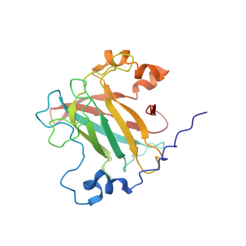 | |
UniProt | |||||
Find proteins for P00436 (Pseudomonas putida) Explore P00436 Go to UniProtKB: P00436 | |||||
Entity Groups | |||||
| Sequence Clusters | 30% Identity50% Identity70% Identity90% Identity95% Identity100% Identity | ||||
| UniProt Group | P00436 | ||||
Sequence AnnotationsExpand | |||||
| |||||
Entity ID: 2 | |||||
|---|---|---|---|---|---|
| Molecule | Chains | Sequence Length | Organism | Details | Image |
| Protocatechuate 3,4-dioxygenase beta chain | D [auth M], E [auth N], F [auth O] | 238 | Pseudomonas putida | Mutation(s): 2 Gene Names: pcaH EC: 1.13.11.3 | 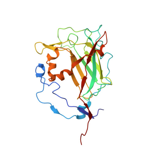 |
UniProt | |||||
Find proteins for P00437 (Pseudomonas putida) Explore P00437 Go to UniProtKB: P00437 | |||||
Entity Groups | |||||
| Sequence Clusters | 30% Identity50% Identity70% Identity90% Identity95% Identity100% Identity | ||||
| UniProt Group | P00437 | ||||
Sequence AnnotationsExpand | |||||
| |||||
| Ligands 6 Unique | |||||
|---|---|---|---|---|---|
| ID | Chains | Name / Formula / InChI Key | 2D Diagram | 3D Interactions | |
| DHB Query on DHB | CA [auth O], S [auth M], Y [auth N] | 3,4-DIHYDROXYBENZOIC ACID C7 H6 O4 YQUVCSBJEUQKSH-UHFFFAOYSA-N |  | ||
| SO4 Query on SO4 | G [auth A] H [auth A] J [auth B] K [auth B] N [auth C] | SULFATE ION O4 S QAOWNCQODCNURD-UHFFFAOYSA-L |  | ||
| GOL Query on GOL | I [auth A], O [auth C], P [auth C] | GLYCEROL C3 H8 O3 PEDCQBHIVMGVHV-UHFFFAOYSA-N |  | ||
| BME Query on BME | L [auth B] M [auth B] Q [auth M] U [auth N] V [auth N] | BETA-MERCAPTOETHANOL C2 H6 O S DGVVWUTYPXICAM-UHFFFAOYSA-N |  | ||
| FE Query on FE | BA [auth O], R [auth M], X [auth N] | FE (III) ION Fe VTLYFUHAOXGGBS-UHFFFAOYSA-N |  | ||
| CL Query on CL | AA [auth O] | CHLORIDE ION Cl VEXZGXHMUGYJMC-UHFFFAOYSA-M |  | ||
| Length ( Å ) | Angle ( ˚ ) |
|---|---|
| a = 128.142 | α = 90 |
| b = 140.831 | β = 90 |
| c = 167.83 | γ = 90 |
| Software Name | Purpose |
|---|---|
| DENZO | data reduction |
| SCALEPACK | data scaling |
| MOLREP | phasing |
| REFMAC | refinement |
| PDB_EXTRACT | data extraction |
| HKL-3000 | data collection |


