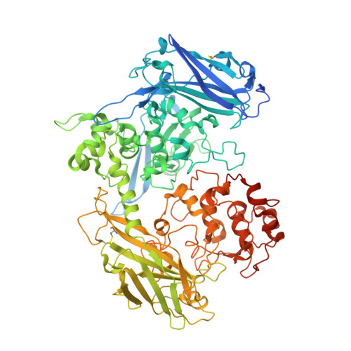
3UFK | pdb_00003ufk

Crystal structure of UndA complexed with Iron Nitrilotriacetate


Experimental Data Snapshot

  • Method: X-RAY DIFFRACTION
  • Resolution: 2.10 Å
  • R-Value Free: 
    0.205 (Depositor), 0.200 (DCC) 
  • R-Value Work: 
    0.155 (Depositor), 0.160 (DCC) 
  • R-Value Observed: 
    0.158 (Depositor) 

wwPDB Validation   3D Report Full Report


Ligand Structure Quality Assessment 


This is version 2.1 of the entry. See complete history


Literature

The Crystal Structure of the Extracellular 11-heme Cytochrome UndA Reveals a Conserved 10-heme Motif and Defined Binding Site for Soluble Iron Chelates.

Edwards, M.J.Hall, A.Shi, L.Fredrickson, J.K.Zachara, J.M.Butt, J.N.Richardson, D.J.Clarke, T.A.

(2012) Structure 20: 1275-1284

  • DOI: https://doi.org/10.1016/j.str.2012.04.016
  • Primary Citation of Related Structures:  
    3UCP, 3UFH, 3UFK

  • PubMed Abstract: 

    Members of the genus Shewanella translocate deca- or undeca-heme cytochromes to the external cell surface thus enabling respiration using extracellular minerals and polynuclear Fe(III) chelates. The high resolution structure of the first undeca-heme outer membrane cytochrome, UndA, reveals a crossed heme chain with four potential electron ingress/egress sites arranged within four domains. Sequence and structural alignment of UndA and the deca-heme MtrF reveals the extra heme of UndA is inserted between MtrF hemes 6 and 7. The remaining UndA hemes can be superposed over the heme chain of the decaheme MtrF, suggesting that a ten heme core is conserved between outer membrane cytochromes. The UndA structure has also been crystallographically resolved in complex with substrates, an Fe(III)-nitrilotriacetate dimer or an Fe(III)-citrate trimer. The structural resolution of these UndA-Fe(III)-chelate complexes provides a rationale for previous kinetic measurements on UndA and other outer membrane cytochromes.


  • Organizational Affiliation
    • Centre for Molecular and Structural Biochemistry, School of Biological Sciences and School of Chemistry, University of East Anglia, Norwich Research Park, Norwich NR4 7TJ, UK.

Macromolecules
Find similar proteins by:  (by identity cutoff)  |  3D Structure
Entity ID: 1
MoleculeChains Sequence LengthOrganismDetailsImage
UndA874Shewanella sp. HRCR_06Mutation(s): 0 
Gene Names: unda
UniProt
Find proteins for F8UWD6 (Shewanella sp. HRCR_06)
Explore F8UWD6 
Go to UniProtKB:  F8UWD6
Entity Groups  
Sequence Clusters30% Identity50% Identity70% Identity90% Identity95% Identity100% Identity
UniProt GroupF8UWD6
Sequence Annotations
Expand
  • Reference Sequence
Small Molecules
Ligands 7 Unique
IDChains Name / Formula / InChI Key2D Diagram3D Interactions
HEC
Query on HEC

Download Ideal Coordinates CCD File 
B [auth A]
C [auth A]
D [auth A]
E [auth A]
F [auth A]
B [auth A],
C [auth A],
D [auth A],
E [auth A],
F [auth A],
G [auth A],
H [auth A],
I [auth A],
J [auth A],
K [auth A],
L [auth A]
HEME C
C34 H34 Fe N4 O4
HXQIYSLZKNYNMH-LJNAALQVSA-N
NTA
Query on NTA

Download Ideal Coordinates CCD File 
S [auth A],
T [auth A],
X [auth A],
Y [auth A]
NITRILOTRIACETIC ACID
C6 H9 N O6
MGFYIUFZLHCRTH-UHFFFAOYSA-N
GOL
Query on GOL

Download Ideal Coordinates CCD File 
AA [auth A],
BA [auth A],
CA [auth A],
Z [auth A]
GLYCEROL
C3 H8 O3
PEDCQBHIVMGVHV-UHFFFAOYSA-N
FE
Query on FE

Download Ideal Coordinates CCD File 
P [auth A],
Q [auth A],
U [auth A],
V [auth A]
FE (III) ION
Fe
VTLYFUHAOXGGBS-UHFFFAOYSA-N
CA
Query on CA

Download Ideal Coordinates CCD File 
M [auth A]CALCIUM ION
Ca
BHPQYMZQTOCNFJ-UHFFFAOYSA-N
MG
Query on MG

Download Ideal Coordinates CCD File 
N [auth A],
O [auth A]
MAGNESIUM ION
Mg
JLVVSXFLKOJNIY-UHFFFAOYSA-N
O
Query on O

Download Ideal Coordinates CCD File 
R [auth A],
W [auth A]
OXYGEN ATOM
O
XLYOFNOQVPJJNP-UHFFFAOYSA-N
Experimental Data & Validation

Experimental Data

  • Method: X-RAY DIFFRACTION
  • Resolution: 2.10 Å
  • R-Value Free:  0.205 (Depositor), 0.200 (DCC) 
  • R-Value Work:  0.155 (Depositor), 0.160 (DCC) 
  • R-Value Observed: 0.158 (Depositor) 
Space Group: P 21 21 21
Unit Cell:
Length ( Å )Angle ( ˚ )
a = 69.5α = 90
b = 105.87β = 90
c = 151.52γ = 90
Software Package:
Software NamePurpose
xia2data scaling
PHASERphasing
REFMACrefinement
xia2data reduction

Structure Validation

View Full Validation Report


Ligand Structure Quality Assessment 


Entry History 

Deposition Data

Revision History  (Full details and data files)

  • Version 1.0: 2012-08-08
    Type: Initial release
  • Version 2.0: 2023-11-15
    Changes: Atomic model, Data collection, Database references, Derived calculations
  • Version 2.1: 2024-11-20
    Changes: Structure summary