Primary Citation of Related Structures: 4Q6D, 4Q6E
PubMed Abstract:
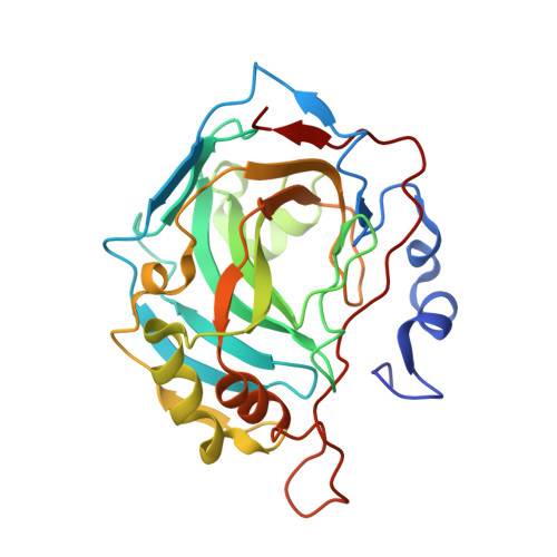
A series of N-aryl-β-alanine derivatives and diazobenzenesulfonamides containing aliphatic rings were designed, synthesized, and their binding to carbonic anhydrases (CA) I, II, VI, VII, XII, and XIII was studied by the fluorescent thermal shift assay and isothermal titration calorimetry. The results showed that 4-substituted diazobenzenesulfonamides were more potent CA binders than N-aryl-β-alanine derivatives. Most of the N-aryl-β-alanine derivatives showed better affinity for CA II while diazobenzenesulfonamides possessed nanomolar affinities towards CA I isozyme. X-ray crystallographic structures showed the modes of binding of both compound groups.
Organizational Affiliation:
Department of Organic Chemistry, Kaunas University of Technology, Kaunas LT-50254, Lithuania. kestutis.rutkauskas@ktu.lt.
Department of Biothermodynamics and Drug Design, Institute of Biotechnology, Vilnius University, Graičiūno 8, Vilnius LT-02241, Lithuania. astzu@ibt.lt.
Department of Organic Chemistry, Kaunas University of Technology, Kaunas LT-50254, Lithuania. ingrida.tumosiene@ktu.lt.
Department of Physical and Inorganic Chemistry, Kaunas University of Technology, Kaunas LT-50254, Lithuania. kristina.kantminiene@ktu.lt.
Institute of Biochemistry, Vilnius University, Mokslininkų 12, Vilnius LT-08862, Lithuania. kazemary@gmail.com.
Department of Biothermodynamics and Drug Design, Institute of Biotechnology, Vilnius University, Graičiūno 8, Vilnius LT-02241, Lithuania. alexeyus1@gmail.com.
Department of Biothermodynamics and Drug Design, Institute of Biotechnology, Vilnius University, Graičiūno 8, Vilnius LT-02241, Lithuania. kazokaite@ibt.lt.
Department of Biothermodynamics and Drug Design, Institute of Biotechnology, Vilnius University, Graičiūno 8, Vilnius LT-02241, Lithuania. morkunaite@ibt.lt.
Department of Biothermodynamics and Drug Design, Institute of Biotechnology, Vilnius University, Graičiūno 8, Vilnius LT-02241, Lithuania. edita.capkauskaite@chf.stud.vu.lt.
Department of Protein-DNA Interactions, Vilnius University Institute of Biotechnology, Graičiūno 8, Vilnius LT-02241, Lithuania. lena@ibt.lt.
Department of Protein-DNA Interactions, Vilnius University Institute of Biotechnology, Graičiūno 8, Vilnius LT-02241, Lithuania. grazulis@ibt.lt.
Department of Organic Chemistry, Kaunas University of Technology, Kaunas LT-50254, Lithuania. zigmuntas.beresnevicius@ktu.lt.
Department of Biothermodynamics and Drug Design, Institute of Biotechnology, Vilnius University, Graičiūno 8, Vilnius LT-02241, Lithuania. daumantas.matulis@bti.vu.lt.