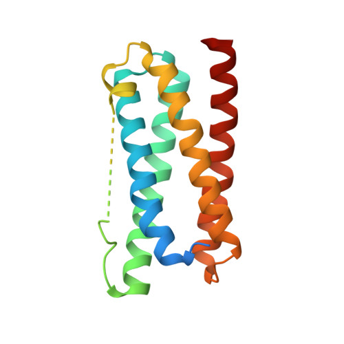
5FFE | pdb_00005ffe

CopM in the Ag-bound form (by soaking)


Experimental Data Snapshot

  • Method: X-RAY DIFFRACTION
  • Resolution: 2.10 Å
  • R-Value Free: 
    0.257 (Depositor), 0.260 (DCC) 
  • R-Value Work: 
    0.214 (Depositor), 0.210 (DCC) 
  • R-Value Observed: 
    0.216 (Depositor) 

Starting Model: experimental
View more details

wwPDB Validation   3D Report Full Report


This is version 1.2 of the entry. See complete history


Literature

Structural basis for copper/silver binding by the Synechocystis metallochaperone CopM.

Zhao, S.Wang, X.Niu, G.Dong, W.Wang, J.Fang, Y.Lin, Y.Liu, L.

(2016) Acta Crystallogr D Struct Biol 72: 997-1005

  • DOI: https://doi.org/10.1107/S2059798316011943
  • Primary Citation of Related Structures:  
    5FEJ, 5FFA, 5FFB, 5FFC, 5FFD, 5FFE

  • PubMed Abstract: 

    Copper homeostasis integrates multiple processes from sensing to storage and efflux out of the cell. CopM is a cyanobacterial metallochaperone, the gene for which is located upstream of a two-component system for copper resistance, but the molecular basis for copper recognition by this four-helical bundle protein is unknown. Here, crystal structures of CopM in apo, copper-bound and silver-bound forms are reported. Monovalent copper/silver ions are buried within the bundle core; divalent copper ions are found on the surface of the bundle. The monovalent copper/silver-binding site is constituted by two consecutive histidines and is conserved in a previously functionally unknown protein family. The structural analyses show two conformational states and suggest that flexibility in the first α-helix is related to the metallochaperone function. These results also reveal functional diversity from a protein family with a simple four-helical fold.


  • Organizational Affiliation
    • Key Laboratory of Photobiology, CAS Center for Excellence in Molecular Plant Sciences, Institute of Botany, Chinese Academy of Sciences, Beijing 100093, People's Republic of China.

Macromolecules
Find similar proteins by:  (by identity cutoff)  |  3D Structure
Entity ID: 1
MoleculeChains Sequence LengthOrganismDetailsImage
CopM
A, B
172Synechocystis sp. PCC 6803Mutation(s): 0 
Gene Names: pcopM
UniProt
Find proteins for Q6YRW5 (Synechocystis sp. (strain ATCC 27184 / PCC 6803 / Kazusa))
Explore Q6YRW5 
Go to UniProtKB:  Q6YRW5
Entity Groups  
Sequence Clusters30% Identity50% Identity70% Identity90% Identity95% Identity100% Identity
UniProt GroupQ6YRW5
Sequence Annotations
Expand
  • Reference Sequence
Small Molecules
Ligands 1 Unique
IDChains Name / Formula / InChI Key2D Diagram3D Interactions
AG
Query on AG

Download Ideal Coordinates CCD File 
C [auth A]
D [auth A]
E [auth A]
F [auth A]
G [auth A]
C [auth A],
D [auth A],
E [auth A],
F [auth A],
G [auth A],
H [auth A],
I [auth B],
J [auth B],
K [auth B],
L [auth B],
M [auth B],
N [auth B]
SILVER ION
Ag
FOIXSVOLVBLSDH-UHFFFAOYSA-N
Experimental Data & Validation

Experimental Data

  • Method: X-RAY DIFFRACTION
  • Resolution: 2.10 Å
  • R-Value Free:  0.257 (Depositor), 0.260 (DCC) 
  • R-Value Work:  0.214 (Depositor), 0.210 (DCC) 
  • R-Value Observed: 0.216 (Depositor) 
Space Group: P 21 21 21
Unit Cell:
Length ( Å )Angle ( ˚ )
a = 53.984α = 90
b = 67.035β = 90
c = 85.614γ = 90
Software Package:
Software NamePurpose
HKL-2000data collection
HKL-2000data scaling
PHASERphasing
PHENIXrefinement
PDB_EXTRACTdata extraction
HKL-2000data reduction
PHASERphasing

Structure Validation

View Full Validation Report


Entry History 

Deposition Data

Revision History  (Full details and data files)

  • Version 1.0: 2016-09-07
    Type: Initial release
  • Version 1.1: 2019-12-25
    Changes: Database references, Derived calculations
  • Version 1.2: 2023-11-08
    Changes: Data collection, Database references, Derived calculations, Refinement description