5M7Y | pdb_00005m7y

Crystal structure of GH125 1,6-alpha-mannosidase mutant from Clostridium perfringens in complex with 1,6-alpha-mannotriose


Experimental Data Snapshot

  • Method: X-RAY DIFFRACTION
  • Resolution: 1.55 Å
  • R-Value Free: 
    0.180 (Depositor), 0.190 (DCC) 
  • R-Value Work: 
    0.147 (Depositor), 0.160 (DCC) 
  • R-Value Observed: 
    0.149 (Depositor) 

wwPDB Validation   3D Report Full Report


This is version 2.1 of the entry. See complete history


Literature

Computational Design of Experiment Unveils the Conformational Reaction Coordinate of GH125 alpha-Mannosidases.

Alonso-Gil, S.Males, A.Fernandes, P.Z.Williams, S.J.Davies, G.J.Rovira, C.

(2017) J Am Chem Soc 139: 1085-1088

  • DOI: https://doi.org/10.1021/jacs.6b11247
  • Primary Citation of Related Structures:  
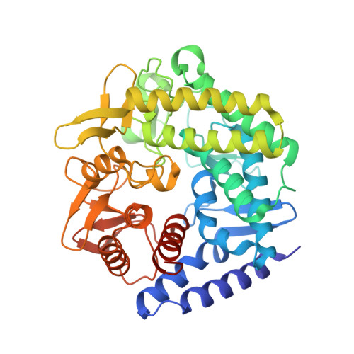
    5M7I, 5M7Y

  • PubMed Abstract: 

    Conformational analysis of enzyme-catalyzed mannoside hydrolysis has revealed two predominant conformational itineraries through B 2,5 or 3 H 4 transition-state (TS) conformations. A prominent unassigned catalytic itinerary is that of exo-1,6-α-mannosidases belonging to CAZy family 125. A published complex of Clostridium perfringens GH125 enzyme with a nonhydrolyzable 1,6-α-thiomannoside substrate mimic bound across the active site revealed an undistorted 4 C 1 conformation and provided no insight into the catalytic pathway of this enzyme. We show through a purely computational approach (QM/MM metadynamics) that sulfur-for-oxygen substitution in the glycosidic linkage fundamentally alters the energetically accessible conformational space of a thiomannoside when bound within the GH125 active site. Modeling of the conformational free energy landscape (FEL) of a thioglycoside strongly favors a mechanistically uninformative 4 C 1 conformation within the GH125 enzyme active site, but the FEL of corresponding O-glycoside substrate reveals a preference for a Michaelis complex in an O S 2 conformation (consistent with catalysis through a B 2,5 TS). This prediction was tested experimentally by determination of the 3D X-ray structure of the pseudo-Michaelis complex of an inactive (D220N) variant of C. perfringens GH125 enzyme in complex with 1,6-α-mannobiose. This complex revealed unambiguous distortion of the -1 subsite mannoside to an O S 2 conformation, matching that predicted by theory and supporting an O S 2 → B 2,5 1 S 5 conformational itinerary for GH125 α-mannosidases. This work highlights the power of the QM/MM approach and identified shortcomings in the use of nonhydrolyzable substrate analogues for conformational analysis of enzyme-bound species.


  • Organizational Affiliation
    • Departament de Química Inorgànica i Orgànica (Secció de Química Orgànica) & Institut de Química Teòrica i Computacional (IQTCUB), Universitat de Barcelona , 08028 Barcelona, Spain.

Macromolecules
Find similar proteins by:  (by identity cutoff)  |  3D Structure
Entity ID: 1
MoleculeChains Sequence LengthOrganismDetailsImage
1,6-alpha-mannosidase435Clostridium perfringens str. 13Mutation(s): 1 
Gene Names: CPE0426
UniProt
Find proteins for Q8XNB2 (Clostridium perfringens (strain 13 / Type A))
Explore Q8XNB2 
Go to UniProtKB:  Q8XNB2
Entity Groups  
Sequence Clusters30% Identity50% Identity70% Identity90% Identity95% Identity100% Identity
UniProt GroupQ8XNB2
Sequence Annotations
Expand
  • Reference Sequence
Oligosaccharides

Help

Entity ID: 2
MoleculeChains Length2D Diagram Glycosylation3D Interactions
alpha-D-mannopyranose-(1-6)-alpha-D-mannopyranose-(1-6)-alpha-D-mannopyranose
B
3N/A
Glycosylation Resources
GlyTouCan:  G85415LL
GlyCosmos:  G85415LL
GlyGen:  G85415LL
Experimental Data & Validation

Experimental Data

  • Method: X-RAY DIFFRACTION
  • Resolution: 1.55 Å
  • R-Value Free:  0.180 (Depositor), 0.190 (DCC) 
  • R-Value Work:  0.147 (Depositor), 0.160 (DCC) 
  • R-Value Observed: 0.149 (Depositor) 
Space Group: P 1 21 1
Unit Cell:
Length ( Å )Angle ( ˚ )
a = 60.414α = 90
b = 44.016β = 96.59
c = 85.079γ = 90
Software Package:
Software NamePurpose
REFMACrefinement
xia2data reduction
Aimlessdata scaling

Structure Validation

View Full Validation Report


Entry History & Funding Information

Deposition Data


Funding OrganizationLocationGrant Number
Biotechnology and Biological Sciences Research CouncilUnited Kingdom--

Revision History  (Full details and data files)

  • Version 1.0: 2016-11-30
    Type: Initial release
  • Version 1.1: 2017-01-11
    Changes: Database references
  • Version 1.2: 2017-02-08
    Changes: Database references
  • Version 1.3: 2017-08-30
    Changes: Author supporting evidence
  • Version 2.0: 2020-07-29
    Type: Remediation
    Reason: Carbohydrate remediation
    Changes: Atomic model, Data collection, Derived calculations, Structure summary
  • Version 2.1: 2024-05-08
    Changes: Data collection, Database references, Derived calculations, Structure summary