5V8E | pdb_00005v8e

Structure of Bacillus cereus PatB1


Experimental Data Snapshot

  • Method: X-RAY DIFFRACTION
  • Resolution: 2.20 Å
  • R-Value Free: 
    0.241 (Depositor), 0.240 (DCC) 
  • R-Value Work: 
    0.199 (Depositor), 0.200 (DCC) 
  • R-Value Observed: 
    0.201 (Depositor) 

wwPDB Validation   3D Report Full Report


This is version 1.3 of the entry. See complete history


Literature

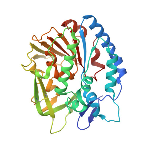
PatB1 is an O-acetyltransferase that decorates secondary cell wall polysaccharides.

Sychantha, D.Little, D.J.Chapman, R.N.Boons, G.J.Robinson, H.Howell, P.L.Clarke, A.J.

(2018) Nat Chem Biol 14: 79-85

  • DOI: https://doi.org/10.1038/nchembio.2509
  • Primary Citation of Related Structures:  
    5V8D, 5V8E

  • PubMed Abstract: 

    O-Acetylation of the secondary cell wall polysaccharides (SCWP) of the Bacillus cereus group of pathogens, which includes Bacillus anthracis, is essential for the proper attachment of surface-layer (S-layer) proteins to their cell walls. Using a variety of pseudosubstrates and a chemically synthesized analog of SCWP, we report here the identification of PatB1 as a SCWP O-acetyltransferase in Bacillus cereus. Additionally, we report the crystal structure of PatB1, which provides detailed insights into the mechanism of this enzyme and defines a novel subfamily of the SGNH family of esterases and lipases. We propose a model for the O-acetylation of SCWP requiring the translocation of acetyl groups from a cytoplasmic source across the plasma membrane by PatA1 and PatA2 for their transfer to SCWP by PatB1.


  • Organizational Affiliation
    • Department of Molecular and Cellular Biology, University of Guelph, Guelph, Ontario, Canada.

Macromolecules
Find similar proteins by:  (by identity cutoff)  |  3D Structure
Entity ID: 1
MoleculeChains Sequence LengthOrganismDetailsImage
Bacillus cereus PatB1
A, B
309Bacillus cereus ATCC 10987Mutation(s): 6 
Gene Names: BCE_0974
UniProt
Find proteins for Q73CU0 (Bacillus cereus (strain ATCC 10987 / NRS 248))
Explore Q73CU0 
Go to UniProtKB:  Q73CU0
Entity Groups  
Sequence Clusters30% Identity50% Identity70% Identity90% Identity95% Identity100% Identity
UniProt GroupQ73CU0
Sequence Annotations
Expand
  • Reference Sequence
Small Molecules
Ligands 3 Unique
IDChains Name / Formula / InChI Key2D Diagram3D Interactions
CIT
Query on CIT

Download Ideal Coordinates CCD File 
C [auth A],
D [auth A],
H [auth B]
CITRIC ACID
C6 H8 O7
KRKNYBCHXYNGOX-UHFFFAOYSA-N
PEG
Query on PEG

Download Ideal Coordinates CCD File 
F [auth A],
G [auth A],
I [auth B]
DI(HYDROXYETHYL)ETHER
C4 H10 O3
MTHSVFCYNBDYFN-UHFFFAOYSA-N
NA
Query on NA

Download Ideal Coordinates CCD File 
E [auth A]SODIUM ION
Na
FKNQFGJONOIPTF-UHFFFAOYSA-N
Experimental Data & Validation

Experimental Data

  • Method: X-RAY DIFFRACTION
  • Resolution: 2.20 Å
  • R-Value Free:  0.241 (Depositor), 0.240 (DCC) 
  • R-Value Work:  0.199 (Depositor), 0.200 (DCC) 
  • R-Value Observed: 0.201 (Depositor) 
Space Group: P 63
Unit Cell:
Length ( Å )Angle ( ˚ )
a = 133.76α = 90
b = 133.76β = 90
c = 87.311γ = 120
Software Package:
Software NamePurpose
PHENIXrefinement
XDSdata reduction
XDSdata scaling
PHASERphasing
PHASERmodel building

Structure Validation

View Full Validation Report


Entry History 

Deposition Data

Revision History  (Full details and data files)

  • Version 1.0: 2017-10-18
    Type: Initial release
  • Version 1.1: 2017-12-06
    Changes: Database references
  • Version 1.2: 2017-12-20
    Changes: Database references
  • Version 1.3: 2024-10-23
    Changes: Data collection, Database references, Structure summary