8IYM | pdb_00008iym

Crystal structure of a protein acetyltransferase, HP0935


Experimental Data Snapshot

  • Method: X-RAY DIFFRACTION
  • Resolution: 2.00 Å
  • R-Value Free: 
    0.246 (Depositor), 0.250 (DCC) 
  • R-Value Work: 
    0.215 (Depositor), 0.220 (DCC) 
  • R-Value Observed: 
    0.217 (Depositor) 

Starting Model: experimental
View more details

wwPDB Validation   3D Report Full Report


This is version 2.1 of the entry. See complete history


Literature

Biochemical and structural characterization of a GNAT superfamily protein acetyltransferase from Helicobacter pylori.

Dadireddy, V.Kumar, A.Kumar, S.Sarma, S.P.Mahanta, P.Ramakumar, S.Desirazu, R.N.

(2025) J Biological Chem 301: 110356-110356

  • DOI: https://doi.org/10.1016/j.jbc.2025.110356
  • Primary Citation of Related Structures:  
    8IYM, 8IYO

  • PubMed Abstract: 

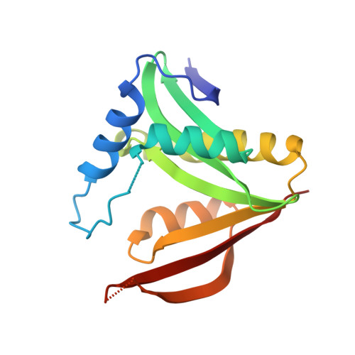
    Helicobacter pylori (H. pylori), a gastric pathogen with high genetic variability and a unique niche, causes peptic ulcers and gastric cancer. Natural transformation contributes to the genetic variability of H. pylori. To date, protein acetylation and the associated acetyltransferase(s) have not been reported in this bacterium. Here, we report protein acetylation in H. pylori and identify a putative protein acetyltransferase, HP0935, capable of acetylating amino acids and proteins, including DNA processing protein A (DprA), which is involved in natural transformation. HP0935 acetylates residue K133 in DprA, which is important for DNA binding, thus is likely to regulate natural transformation. We determined the crystal structures of HP0935 in its apo form and in complex with acetyl-coenzyme A (ACO) to 2.00 Å and 2.40 Å resolution, respectively. Structural analysis revealed a conformational change in the substrate-binding loops, α1-α2 and β6-β7, upon ACO binding. The structural comparison showed that HP0935 differs from other protein acetyltransferases in the length and orientation of these loops. Molecular dynamics simulation data suggest that these loops are highly dynamic, and ACO binding could affect their dynamics. Given that several proteins may undergo acetylation in H. pylori and the fact that HP0935 is the only known protein acetyltransferase, the loop dynamics are likely to facilitate the acceptance of multiple substrates by HP0935. Structure-based mutational analysis showed that no general base is required for the enzymatic activity. However, a conserved catalytic water molecule at the active site is likely to serve the purpose. Furthermore, the general acid Y127 is essential for enzymatic activity.


  • Organizational Affiliation
    • Department of Physics, Indian Institute of Science, Bangalore, India.

Macromolecules
Find similar proteins by:  (by identity cutoff)  |  3D Structure
Entity ID: 1
MoleculeChains Sequence LengthOrganismDetailsImage
N-acetyltransferase domain-containing protein
A, B, C
163Helicobacter pylori 26695Mutation(s): 1 
Gene Names: HP_0935
UniProt
Find proteins for O25589 (Helicobacter pylori (strain ATCC 700392 / 26695))
Explore O25589 
Go to UniProtKB:  O25589
Entity Groups  
Sequence Clusters30% Identity50% Identity70% Identity90% Identity95% Identity100% Identity
UniProt GroupO25589
Sequence Annotations
Expand
  • Reference Sequence
Small Molecules
Ligands 3 Unique
IDChains Name / Formula / InChI Key2D Diagram3D Interactions
EDO
Query on EDO

Download Ideal Coordinates CCD File 
F [auth A],
I [auth B],
P [auth C]
1,2-ETHANEDIOL
C2 H6 O2
LYCAIKOWRPUZTN-UHFFFAOYSA-N
SCN
Query on SCN

Download Ideal Coordinates CCD File 
G [auth A]
H [auth A]
J [auth B]
K [auth B]
L [auth B]
G [auth A],
H [auth A],
J [auth B],
K [auth B],
L [auth B],
M [auth B],
N [auth B],
Q [auth C],
R [auth C]
THIOCYANATE ION
C N S
ZMZDMBWJUHKJPS-UHFFFAOYSA-M
K
Query on K

Download Ideal Coordinates CCD File 
D [auth A],
E [auth A],
O [auth C]
POTASSIUM ION
K
NPYPAHLBTDXSSS-UHFFFAOYSA-N
Experimental Data & Validation

Experimental Data

  • Method: X-RAY DIFFRACTION
  • Resolution: 2.00 Å
  • R-Value Free:  0.246 (Depositor), 0.250 (DCC) 
  • R-Value Work:  0.215 (Depositor), 0.220 (DCC) 
  • R-Value Observed: 0.217 (Depositor) 
Space Group: C 1 2 1
Unit Cell:
Length ( Å )Angle ( ˚ )
a = 117.856α = 90
b = 66.17β = 91.42
c = 70.904γ = 90
Software Package:
Software NamePurpose
REFMACrefinement
Aimlessdata scaling
PHASERphasing
PDB_EXTRACTdata extraction
PROTEUM PLUSdata reduction

Structure Validation

View Full Validation Report


Entry History & Funding Information

Deposition Data


Funding OrganizationLocationGrant Number
Not funded--

Revision History  (Full details and data files)

  • Version 1.0: 2024-04-10
    Type: Initial release
  • Version 2.0: 2024-05-08
    Type: Coordinate replacement
    Reason: Real space R-factor
    Changes: Atomic model, Data collection, Derived calculations, Refinement description, Structure summary
  • Version 2.1: 2025-07-30
    Changes: Database references, Structure summary