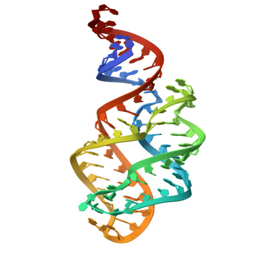
Crystal structures reveal the distinct features of the 2'-dG-III riboswitch in the purine riboswitch family.
Chen, K., Liao, W., Wang, J., Ren, Y., Lu, Z., Peng, X., Wang, J., Huang, L.(2025) Nucleic Acids Res 53
- PubMed: 40694847
- DOI: https://doi.org/10.1093/nar/gkaf702
- Primary Citation of Related Structures:
8KEB, 8KED, 8KHH - PubMed Abstract:
Purine riboswitches, located in the 5'-untranslated regions of bacterial messenger RNA, regulate gene expression by sensing purines and their derivatives. A class of the guanine-I riboswitch variants was recently reported to be the third class of 2'-deoxyguanosine (2'-dG) riboswitch termed 2'-dG-III riboswitch. Here we present the crystal structures of the 2'-dG-III riboswitch bound with 2'-dG, guanosine, or guanine. Despite similarities in secondary and overall structures to other purine riboswitches, the 2'-dG-III riboswitch exhibits unique features in its loop-loop interaction, three-way junction, and ligand binding. The 2'-dG-III riboswitch exhibits a tuning fork-like structure with a unique six-tiered interaction within the three-way junction. The second, third, and fourth tiers form the ligand-binding pocket, with a consistent binding mode for the guanine moiety of all three ligands. Structural and biochemical analyses reveal detailed interactions between the 2'-dG-III riboswitch and different ligands, providing insights into its regulatory mechanisms in purine metabolism.
- Guangdong Provincial Key Laboratory of Malignant Tumor Epigenetics and Gene Regulation, Guangdong-Hong Kong Joint Laboratory for RNA Medicine, Medical Research Center, Sun Yat-Sen Memorial Hospital, Sun Yat-Sen University, Guangzhou 510120, China.
Organizational Affiliation: