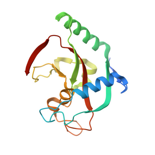
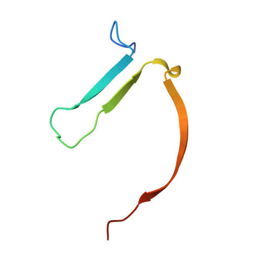
8B6M | pdb_00008b6m

Tankyrase 2 in complex with an inhibitor


Experimental Data Snapshot

  • Method: X-RAY DIFFRACTION
  • Resolution: 1.60 Å
  • R-Value Free: 
    0.219 (Depositor), 0.230 (DCC) 
  • R-Value Work: 
    0.185 (Depositor), 0.200 (DCC) 

Starting Model: experimental
View more details

wwPDB Validation   3D Report Full Report


Ligand Structure Quality Assessment 


This is version 1.1 of the entry. See complete history


Literature

Deconstruction of Dual-Site Tankyrase Inhibitors Provides Insights into Binding Energetics and Suggests Critical Hotspots for Ligand Optimization.

Sowa, S.T.Kucukdisli, M.Mostinski, Y.Schaller, D.A.Vinagreiro, C.S.Cirillo, D.Bosetti, C.Brinch, S.A.van Laar, K.Wegert, A.Leenders, R.G.G.Krauss, S.Waaler, J.Volkamer, A.Lehtio, L.Nazare, M.

(2025) J Med Chem 68: 7263-7279

  • DOI: https://doi.org/10.1021/acs.jmedchem.4c02845
  • Primary Citation of Related Structures:  
    7OJO, 8B6M

  • PubMed Abstract: 

    Designing inhibitors is a complex task that requires a deep understanding of protein-ligand interactions and their dynamics. Ligands often interact with multiple binding subsites, with noncovalent interactions affecting binding affinity. Conformational changes and plasticity of both, the ligand and the protein influence binding energetics. We investigated the tankyrase ADP-ribosyltransferase as a promising drug target regulating many cellular pathways. Despite the existence of diverse tankyrase inhibitors, their binding energetics and contributions of flexible cryptic subpockets to binding affinity remain elusive. To examine these aspects, we deconstructed inhibitors to key fragments, dissected their energetic contribution to the affinity, and determined their binding mode by X-ray crystallography. Varying ligand efficiencies of the deconstructed, pocket-binding fragments revealed the cryptic nature of subpockets. These insights enabled us to redesign inhibitors with novel linkers, the observed key area for optimization, increasing the potency in enzymatic and cell-based assays by 7.5-fold and 6.2-fold compared to the parent ligand.


  • Organizational Affiliation
    • Faculty for Biochemistry and Molecular Medicine & Biocenter Oulu, University of Oulu, Aapistie 7, 90220 Oulu, Finland.

Macromolecules
Find similar proteins by:  (by identity cutoff)  |  3D Structure
Entity ID: 1
MoleculeChains Sequence LengthOrganismDetailsImage
Poly [ADP-ribose] polymerase tankyrase-2
A, C
171Homo sapiensMutation(s): 0 
Gene Names: TNKS2PARP5BTANK2TNKL
EC: 2.4.2.30 (PDB Primary Data), 2.4.2 (PDB Primary Data)
UniProt & NIH Common Fund Data Resources
Find proteins for Q9H2K2 (Homo sapiens)
Explore Q9H2K2 
Go to UniProtKB:  Q9H2K2
PHAROS:  Q9H2K2
GTEx:  ENSG00000107854 
Entity Groups  
Sequence Clusters30% Identity50% Identity70% Identity90% Identity95% Identity100% Identity
UniProt GroupQ9H2K2
Sequence Annotations
Expand
  • Reference Sequence
Find similar proteins by:  (by identity cutoff)  |  3D Structure
Entity ID: 2
MoleculeChains Sequence LengthOrganismDetailsImage
Poly [ADP-ribose] polymerase tankyrase-2
B, D
48Homo sapiensMutation(s): 0 
Gene Names: TNKS2PARP5BTANK2TNKL
EC: 2.4.2.30 (PDB Primary Data), 2.4.2 (PDB Primary Data)
UniProt & NIH Common Fund Data Resources
Find proteins for Q9H2K2 (Homo sapiens)
Explore Q9H2K2 
Go to UniProtKB:  Q9H2K2
PHAROS:  Q9H2K2
GTEx:  ENSG00000107854 
Entity Groups  
Sequence Clusters30% Identity50% Identity70% Identity90% Identity95% Identity100% Identity
UniProt GroupQ9H2K2
Sequence Annotations
Expand
  • Reference Sequence
Small Molecules
Ligands 4 Unique
IDChains Name / Formula / InChI Key2D Diagram3D Interactions
OY6 (Subject of Investigation/LOI)
Query on OY6

Download Ideal Coordinates CCD File 
E [auth A],
K [auth C]
~{N}-(2-methoxyphenyl)-4-[[2-(4-oxidanylidene-3~{H}-quinazolin-2-yl)ethyl-(thiophen-2-ylmethyl)carbamoyl]amino]benzamide
C30 H27 N5 O4 S
CHCNDJNLIDGUFS-UHFFFAOYSA-N
SO4
Query on SO4

Download Ideal Coordinates CCD File 
F [auth A],
G [auth A],
L [auth C],
M [auth D]
SULFATE ION
O4 S
QAOWNCQODCNURD-UHFFFAOYSA-L
GOL
Query on GOL

Download Ideal Coordinates CCD File 
I [auth B]GLYCEROL
C3 H8 O3
PEDCQBHIVMGVHV-UHFFFAOYSA-N
ZN
Query on ZN

Download Ideal Coordinates CCD File 
H [auth A],
J [auth C]
ZINC ION
Zn
PTFCDOFLOPIGGS-UHFFFAOYSA-N
Experimental Data & Validation

Experimental Data

  • Method: X-RAY DIFFRACTION
  • Resolution: 1.60 Å
  • R-Value Free:  0.219 (Depositor), 0.230 (DCC) 
  • R-Value Work:  0.185 (Depositor), 0.200 (DCC) 
Space Group: C 2 2 21
Unit Cell:
Length ( Å )Angle ( ˚ )
a = 90.57α = 90
b = 97.37β = 90
c = 119.24γ = 90
Software Package:
Software NamePurpose
REFMACrefinement
XDSdata reduction
XSCALEdata scaling
PHASERphasing

Structure Validation

View Full Validation Report


Ligand Structure Quality Assessment 


Entry History & Funding Information

Deposition Data


Funding OrganizationLocationGrant Number
Jane and Aatos Erkko FoundationFinland--

Revision History  (Full details and data files)

  • Version 1.0: 2023-10-11
    Type: Initial release
  • Version 1.1: 2025-04-30
    Changes: Database references, Structure summary