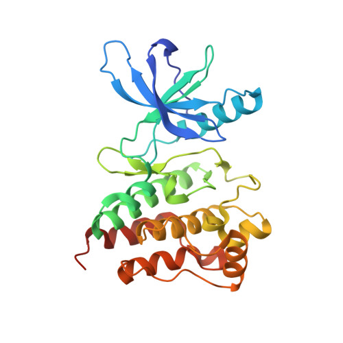
Crystal structure of the kinase domain of a receptor tyrosine kinase from a choanoflagellate, Monosiga brevicollis.
Bajaj, T., Kuriyan, J., Gee, C.L.(2023) PLoS One 18: e0276413-e0276413
- PubMed: 37310965
- DOI: https://doi.org/10.1371/journal.pone.0276413
- Primary Citation of Related Structures:
8E4T - PubMed Abstract:
Genomic analysis of the unicellular choanoflagellate, Monosiga brevicollis (MB), revealed the remarkable presence of cell signaling and adhesion protein domains that are characteristically associated with metazoans. Strikingly, receptor tyrosine kinases, one of the most critical elements of signal transduction and communication in metazoans, are present in choanoflagellates. We determined the crystal structure at 1.95 Å resolution of the kinase domain of the M. brevicollis receptor tyrosine kinase C8 (RTKC8, a member of the choanoflagellate receptor tyrosine kinase C family) bound to the kinase inhibitor staurospaurine. The chonanoflagellate kinase domain is closely related in sequence to mammalian tyrosine kinases (~ 40% sequence identity to the human Ephrin kinase domain EphA3) and, as expected, has the canonical protein kinase fold. The kinase is structurally most similar to human Ephrin (EphA5), even though the extracellular sensor domain is completely different from that of Ephrin. The RTKC8 kinase domain is in an active conformation, with two staurosporine molecules bound to the kinase, one at the active site and another at the peptide-substrate binding site. To our knowledge this is the first example of staurospaurine binding in the Aurora A activation segment (AAS). We also show that the RTKC8 kinase domain can phosphorylate tyrosine residues in peptides from its C-terminal tail segment which is presumably the mechanism by which it transmits the extracellular stimuli to alter cellular function.
- Graduate Program in Comparative Biochemistry, University of California, Berkeley, Berkeley, California, United States of America.
Organizational Affiliation: