8JNZ | pdb_00008jnz

Human ADP-ribosyltransferase 1 (PARP1) catalytic domain bound to a pyrazolopyrimidine carboxamide inhibitor


Experimental Data Snapshot

  • Method: X-RAY DIFFRACTION
  • Resolution: 2.84 Å
  • R-Value Free: 
    0.278 (Depositor), 0.280 (DCC) 
  • R-Value Work: 
    0.219 (Depositor), 0.230 (DCC) 
  • R-Value Observed: 
    0.222 (Depositor) 

Starting Model: experimental
View more details

wwPDB Validation   3D Report Full Report


Ligand Structure Quality Assessment 


This is version 1.1 of the entry. See complete history


Literature

Employing a Highly Potent Fluorescence Probe to Discover a PARP-1/2 Binder and the Complex Structures Analysis.

Wang, X.Wang, C.Li, J.Zhou, J.Xu, B.

(2025) ChemMedChem : e202500168-e202500168

  • DOI: https://doi.org/10.1002/cmdc.202500168
  • Primary Citation of Related Structures:  
    8JNY, 8JNZ

  • PubMed Abstract: 

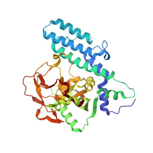
    Poly (ADP-ribose) polymerases-1/2(PARP-1/2) has been identified as important anti-tumor drug targets, and the development of PARP-1/2 inhibitors featuring novel structures is still a promising strategy for cancer treatments. In this work, a highly potent PARP-1/2 probe with a quinazolinone scaffold was designed and synthesized, showing dissociation constants (Kd) of 2.07 nM and 1.6 nM towards catPARP-1 and catPARP-2SE. By employing this probe to screen an in-house compound library, compound A bearing a pyrazolo[1,5-a]pyrimidine-3-carboxamide scaffold was disclosed as a structurally novel PARP-1/2 binder, which had dissociation constants of 5.6 μM and 7.9 μM for catPARP-1 and catPARP-2SE in the Isothermal Titration Calorimetry (ITC) assay. Moreover, the crystal structures of compound A in complex with PARP-1 and PARP-2 catalytic domains were solved to reveal the binding modes of this compound, and these two complex structures were analyzed with IGMH method at GFN2-xTB and B3LYP levels. Interestingly, this compound presented significant differences in binding modes within PARP-1 and PARP-2. These results could provide a structural basis for the discovery of novel PARP-1 or PARP-2 selective inhibitors by taking compound A as a template structure.


  • Organizational Affiliation
    • Institute of Material Medical: Chinese Academy of Medical Sciences & Peking Union Medical College Institute of Materia Medica, synthetic medicinal chemistry, CHINA.

Macromolecules
Find similar proteins by:  (by identity cutoff)  |  3D Structure
Entity ID: 1
MoleculeChains Sequence LengthOrganismDetailsImage
Poly [ADP-ribose] polymerase 1, processed C-terminus
A, B
354Homo sapiensMutation(s): 1 
Gene Names: PARP1ADPRTPPOL
EC: 2.4.2 (UniProt), 2.4.2.30 (UniProt)
UniProt & NIH Common Fund Data Resources
Find proteins for P09874 (Homo sapiens)
Explore P09874 
Go to UniProtKB:  P09874
PHAROS:  P09874
GTEx:  ENSG00000143799 
Entity Groups  
Sequence Clusters30% Identity50% Identity70% Identity90% Identity95% Identity100% Identity
UniProt GroupP09874
Sequence Annotations
Expand
  • Reference Sequence
Experimental Data & Validation

Experimental Data

  • Method: X-RAY DIFFRACTION
  • Resolution: 2.84 Å
  • R-Value Free:  0.278 (Depositor), 0.280 (DCC) 
  • R-Value Work:  0.219 (Depositor), 0.230 (DCC) 
  • R-Value Observed: 0.222 (Depositor) 
Space Group: P 21 21 21
Unit Cell:
Length ( Å )Angle ( ˚ )
a = 48.516α = 90
b = 91.815β = 90
c = 162.941γ = 90
Software Package:
Software NamePurpose
PHENIXrefinement
autoPROCdata processing
Aimlessdata scaling
MOLREPphasing

Structure Validation

View Full Validation Report


Ligand Structure Quality Assessment 


Entry History & Funding Information

Deposition Data


Funding OrganizationLocationGrant Number
National Natural Science Foundation of China (NSFC)China82003657
National Natural Science Foundation of China (NSFC)China81673300
National Natural Science Foundation of China (NSFC)China82173693

Revision History  (Full details and data files)

  • Version 1.0: 2024-06-12
    Type: Initial release
  • Version 1.1: 2025-05-14
    Changes: Database references, Structure summary