8KDL | pdb_00008kdl

Crystal structure of LmbF in complex with PLP


Experimental Data Snapshot

  • Method: X-RAY DIFFRACTION
  • Resolution: 1.70 Å
  • R-Value Free: 
    0.204 (Depositor), 0.200 (DCC) 
  • R-Value Work: 
    0.165 (Depositor), 0.160 (DCC) 
  • R-Value Observed: 
    0.166 (Depositor) 

Starting Model: in silico
View more details

wwPDB Validation   3D Report Full Report


This is version 1.1 of the entry. See complete history


Literature

Molecular basis for the diversification of lincosamide biosynthesis by pyridoxal phosphate-dependent enzymes.

Mori, T.Moriwaki, Y.Sakurada, K.Lyu, S.Kadlcik, S.Janata, J.Mazumdar, A.Koberska, M.Terada, T.Kamenik, Z.Abe, I.

(2025) Nat Chem 17: 256-264

  • DOI: https://doi.org/10.1038/s41557-024-01687-7
  • Primary Citation of Related Structures:  
    8KDK, 8KDL

  • PubMed Abstract: 

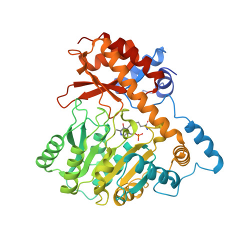
    The biosynthesis of the lincosamide antibiotics lincomycin A and celesticetin involves the pyridoxal-5'-phosphate (PLP)-dependent enzymes LmbF and CcbF, which are responsible for bifurcation of the biosynthetic pathways. Despite recognizing the same S-glycosyl-L-cysteine structure of the substrates, LmbF catalyses thiol formation through β-elimination, whereas CcbF produces S-acetaldehyde through decarboxylation-coupled oxidative deamination. The structural basis for the diversification mechanism remains largely unexplored. Here we conduct structure-function analyses of LmbF and CcbF. X-ray crystal structures, docking and molecular dynamics simulations reveal that active-site aromatic residues play important roles in controlling the substrate binding mode and the reaction outcome. Furthermore, the reaction selectivity and oxygen-utilization of LmbF and CcbF were rationally engineered through structure- and calculation-based mutagenesis. Thus, the catalytic function of CcbF was switched to that of LmbF, and, remarkably, both LmbF and CcbF variants gained the oxidative-amidation activity to produce an unnatural S-acetamide derivative of lincosamide.


  • Organizational Affiliation
    • Graduate School of Pharmaceutical Sciences, The University of Tokyo, Tokyo, Japan. tmori@mol.f.u-tokyo.ac.jp.

Macromolecules
Find similar proteins by:  (by identity cutoff)  |  3D Structure
Entity ID: 1
MoleculeChains Sequence LengthOrganismDetailsImage
Aminotransferase class I/II-fold pyridoxal phosphate-dependent enzyme
A, B
437Streptomyces lincolnensisMutation(s): 0 
Gene Names: GJU35_01420
UniProt
Find proteins for A9Y8R7 (Streptomyces lincolnensis)
Explore A9Y8R7 
Go to UniProtKB:  A9Y8R7
Entity Groups  
Sequence Clusters30% Identity50% Identity70% Identity90% Identity95% Identity100% Identity
UniProt GroupA9Y8R7
Sequence Annotations
Expand
  • Reference Sequence
Small Molecules
Modified Residues  1 Unique
IDChains TypeFormula2D DiagramParent
LLP
Query on LLP
A, B
L-PEPTIDE LINKINGC14 H22 N3 O7 PLYS
Experimental Data & Validation

Experimental Data

  • Method: X-RAY DIFFRACTION
  • Resolution: 1.70 Å
  • R-Value Free:  0.204 (Depositor), 0.200 (DCC) 
  • R-Value Work:  0.165 (Depositor), 0.160 (DCC) 
  • R-Value Observed: 0.166 (Depositor) 
Space Group: P 1 21 1
Unit Cell:
Length ( Å )Angle ( ˚ )
a = 67.277α = 90
b = 74.017β = 93.183
c = 74.843γ = 90
Software Package:
Software NamePurpose
PHENIXrefinement
XDSdata reduction
Aimlessdata scaling
PHASERphasing

Structure Validation

View Full Validation Report


Entry History & Funding Information

Deposition Data


Funding OrganizationLocationGrant Number
Not funded--

Revision History  (Full details and data files)

  • Version 1.0: 2024-08-14
    Type: Initial release
  • Version 1.1: 2025-02-26
    Changes: Database references, Structure summary