8SW8 | pdb_00008sw8

Crystal Structure of HaloTag7 bound to JF669-HaloTag ligand


Experimental Data Snapshot

  • Method: X-RAY DIFFRACTION
  • Resolution: 1.90 Å
  • R-Value Free: 
    0.246 (Depositor), 0.260 (DCC) 
  • R-Value Work: 
    0.194 (Depositor), 0.210 (DCC) 

Starting Model: experimental
View more details

wwPDB Validation   3D Report Full Report


Ligand Structure Quality Assessment 


This is version 1.2 of the entry. See complete history


Literature

A modular chemigenetic calcium indicator for multiplexed in vivo functional imaging.

Farrants, H.Shuai, Y.Lemon, W.C.Monroy Hernandez, C.Zhang, D.Yang, S.Patel, R.Qiao, G.Frei, M.S.Plutkis, S.E.Grimm, J.B.Hanson, T.L.Tomaska, F.Turner, G.C.Stringer, C.Keller, P.J.Beyene, A.G.Chen, Y.Liang, Y.Lavis, L.D.Schreiter, E.R.

(2024) Nat Methods 21: 1916-1925

  • DOI: https://doi.org/10.1038/s41592-024-02411-6
  • Primary Citation of Related Structures:  
    8SW8

  • PubMed Abstract: 

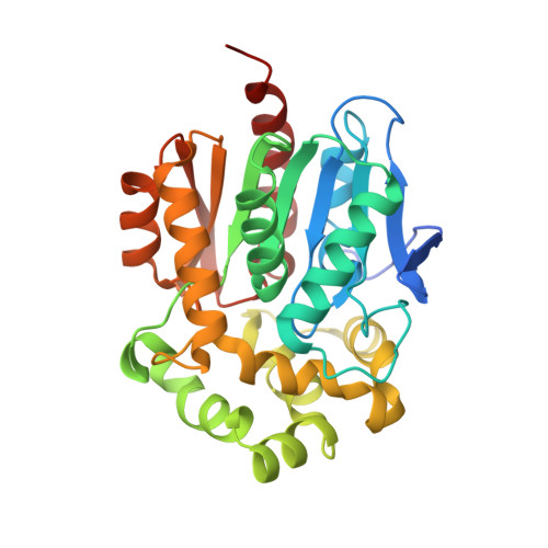
    Genetically encoded fluorescent calcium indicators allow cellular-resolution recording of physiology. However, bright, genetically targetable indicators that can be multiplexed with existing tools in vivo are needed for simultaneous imaging of multiple signals. Here we describe WHaloCaMP, a modular chemigenetic calcium indicator built from bright dye-ligands and protein sensor domains. Fluorescence change in WHaloCaMP results from reversible quenching of the bound dye via a strategically placed tryptophan. WHaloCaMP is compatible with rhodamine dye-ligands that fluoresce from green to near-infrared, including several that efficiently label the brain in animals. When bound to a near-infrared dye-ligand, WHaloCaMP shows a 7× increase in fluorescence intensity and a 2.1-ns increase in fluorescence lifetime upon calcium binding. We use WHaloCaMP1a to image Ca 2+ responses in vivo in flies and mice, to perform three-color multiplexed functional imaging of hundreds of neurons and astrocytes in zebrafish larvae and to quantify Ca 2+ concentration using fluorescence lifetime imaging microscopy (FLIM).


  • Organizational Affiliation
    • Janelia Research Campus, Howard Hughes Medical Institute, Ashburn, VA, USA. farrantsh@janelia.hhmi.org.

Macromolecules
Find similar proteins by:  (by identity cutoff)  |  3D Structure
Entity ID: 1
MoleculeChains Sequence LengthOrganismDetailsImage
Haloalkane dehalogenaseA [auth AAA]304Rhodococcus sp. (in: high G+C Gram-positive bacteria)Mutation(s): 21 
Gene Names: dhaA
EC: 3.8.1.5
UniProt
Find proteins for P0A3G3 (Rhodococcus sp)
Explore P0A3G3 
Go to UniProtKB:  P0A3G3
Entity Groups  
Sequence Clusters30% Identity50% Identity70% Identity90% Identity95% Identity100% Identity
UniProt GroupP0A3G3
Sequence Annotations
Expand
  • Reference Sequence
Small Molecules
Ligands 2 Unique
IDChains Name / Formula / InChI Key2D Diagram3D Interactions
XSR (Subject of Investigation/LOI)
Query on XSR

Download Ideal Coordinates CCD File 
B [auth AAA]1-[(4aM,10P)-7-(azetidin-1-yl)-10-[2-carboxy-3,4,6-trifluoro-5-({2-[2-(hexyloxy)ethoxy]ethyl}carbamoyl)phenyl]-5,5-dimethyldibenzo[b,e]silin-3(5H)-ylidene]azetidin-1-ium
C39 H47 F3 N3 O5 Si
ZQPHKFANJCSITK-UHFFFAOYSA-O
CL
Query on CL

Download Ideal Coordinates CCD File 
C [auth AAA]CHLORIDE ION
Cl
VEXZGXHMUGYJMC-UHFFFAOYSA-M
Experimental Data & Validation

Experimental Data

  • Method: X-RAY DIFFRACTION
  • Resolution: 1.90 Å
  • R-Value Free:  0.246 (Depositor), 0.260 (DCC) 
  • R-Value Work:  0.194 (Depositor), 0.210 (DCC) 
Space Group: P 43 21 2
Unit Cell:
Length ( Å )Angle ( ˚ )
a = 62.741α = 90
b = 62.741β = 90
c = 163.548γ = 90
Software Package:
Software NamePurpose
REFMACrefinement
xia2data reduction
xia2data scaling
PHASERphasing

Structure Validation

View Full Validation Report


Ligand Structure Quality Assessment 


Entry History & Funding Information

Deposition Data


Funding OrganizationLocationGrant Number
Howard Hughes Medical Institute (HHMI)United States--

Revision History  (Full details and data files)

  • Version 1.0: 2024-06-05
    Type: Initial release
  • Version 1.1: 2024-10-16
    Changes: Database references, Structure summary
  • Version 1.2: 2024-10-23
    Changes: Database references