8TQ6 | pdb_00008tq6

Crystal structure of Fab.B1.23.2 in complex with MHC-I (HLA-B*44:05)


Experimental Data Snapshot

  • Method: X-RAY DIFFRACTION
  • Resolution: 3.20 Å
  • R-Value Free: 
    0.268 (Depositor), 0.275 (DCC) 
  • R-Value Work: 
    0.240 (Depositor), 0.242 (DCC) 
  • R-Value Observed: 
    0.253 (Depositor) 

Starting Model: experimental
View more details

wwPDB Validation   3D Report Full Report


This is version 1.1 of the entry. See complete history


Literature

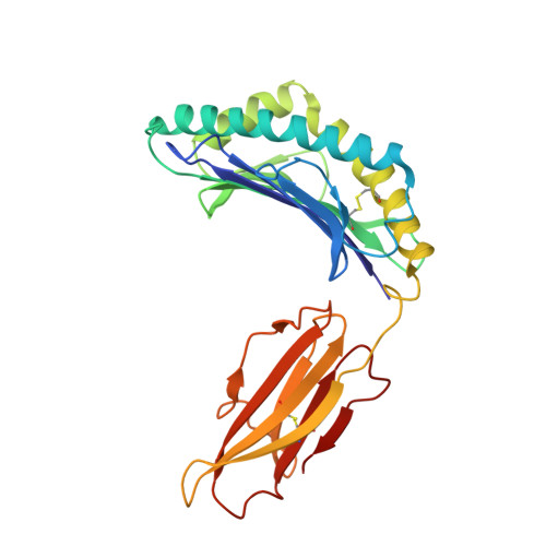
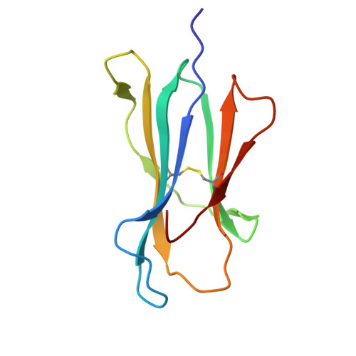
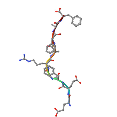
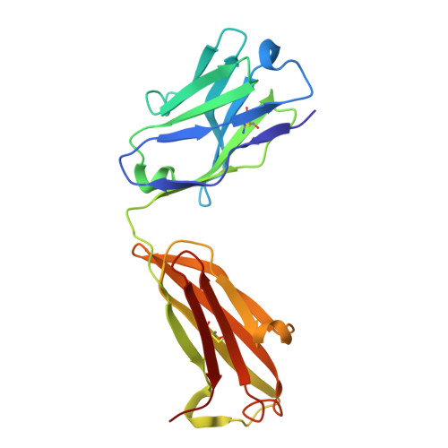
Structural mechanism of anti-MHC-I antibody blocking of inhibitory NK cell receptors in tumor immunity.

Jiang, J.Panda, A.K.Natarajan, K.Lei, H.Sharma, S.Boyd, L.F.Towler, R.R.Chempati, S.Ahmad, J.Morton, A.J.Lang, Z.C.Sun, Y.Sgourakis, N.Meier-Schellersheim, M.Huang, R.K.Shevach, E.M.Margulies, D.H.

(2026) Commun Biol 

  • DOI: https://doi.org/10.1038/s42003-026-09641-8
  • Primary Citation of Related Structures:  
    8TQ6, 9D72, 9D73, 9D74, 9OA9

  • PubMed Abstract: 

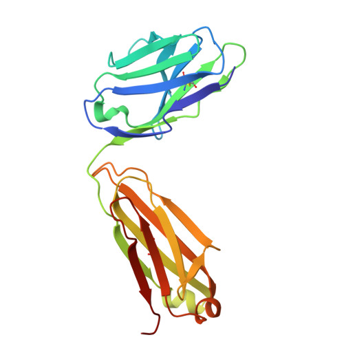
    Anti-major histocompatibility complex class I (MHC-I) mAbs can stimulate immune responses to tumors and infections by blocking suppressive signals delivered via various immune inhibitory receptors. To understand such functions, we determined the structure of a highly cross-reactive anti-human MHC-I mAb, B1.23.2, in complex with the MHC-I molecule HLA-B*44:05 by both cryo-electron microscopy (cryo-EM) and X-ray crystallography. Structural models determined by the two methods were essentially identical revealing that B1.23.2 binds a conserved region on the α2 1 helix that overlaps the killer immunoglobulin-like receptor (KIR) binding site. Structural comparison to KIR/HLA complexes reveals a mechanism by which B1.23.2 blocks inhibitory receptor interactions, leading to natural killer (NK) cell activation. B1.23.2 treatment of the human KLM-1 pancreatic cancer model in humanized (NSG-IL15) mice provides evidence of suppression of tumor growth. Such anti-MHC-I mAb that block inhibitory KIR/HLA interactions may prove useful for tumor immunotherapy.


  • Organizational Affiliation
    • Molecular Biology Section, Laboratory of Immune System Biology, National Institute of Allergy and Infectious Diseases, National Institutes of Health, Bethesda, MD, USA. jiangji@niaid.nih.gov.

Macromolecules
Find similar proteins by:  (by identity cutoff)  |  3D Structure
Entity ID: 1
MoleculeChains Sequence LengthOrganismDetailsImage
HLA class I histocompatibility antigen B alpha chain (HLA-B*44:05)
A, C
274Homo sapiensMutation(s): 0 
Gene Names: HLA-B
UniProt
Find proteins for Q860B7 (Homo sapiens)
Explore Q860B7 
Go to UniProtKB:  Q860B7
Entity Groups  
Sequence Clusters30% Identity50% Identity70% Identity90% Identity95% Identity100% Identity
UniProt GroupQ860B7
Sequence Annotations
Expand
  • Reference Sequence
Find similar proteins by:  (by identity cutoff)  |  3D Structure
Entity ID: 2
MoleculeChains Sequence LengthOrganismDetailsImage
Beta-2-microglobulin
B, D
100Homo sapiensMutation(s): 0 
Gene Names: B2MCDABP0092HDCMA22P
UniProt & NIH Common Fund Data Resources
Find proteins for P61769 (Homo sapiens)
Explore P61769 
Go to UniProtKB:  P61769
PHAROS:  P61769
GTEx:  ENSG00000166710 
Entity Groups  
Sequence Clusters30% Identity50% Identity70% Identity90% Identity95% Identity100% Identity
UniProt GroupP61769
Sequence Annotations
Expand
  • Reference Sequence

Find similar proteins by:  Sequence   |   3D Structure  

Entity ID: 3
MoleculeChains Sequence LengthOrganismDetailsImage
MHC class II antigen peptideE,
J [auth P]
9synthetic constructMutation(s): 0 
UniProt
Find proteins for Q9TQB0 (Homo sapiens)
Explore Q9TQB0 
Go to UniProtKB:  Q9TQB0
Entity Groups  
UniProt GroupQ9TQB0
Sequence Annotations
Expand
  • Reference Sequence
Find similar proteins by:  (by identity cutoff)  |  3D Structure
Entity ID: 4
MoleculeChains Sequence LengthOrganismDetailsImage
Fab B1.23.2 Heavy Chain
F, H
213Mus musculusMutation(s): 0 
Entity Groups  
Sequence Clusters30% Identity50% Identity70% Identity90% Identity95% Identity100% Identity
Sequence Annotations
Expand
  • Reference Sequence
Find similar proteins by:  (by identity cutoff)  |  3D Structure
Entity ID: 5
MoleculeChains Sequence LengthOrganismDetailsImage
Fab B1.23.2 Light ChainG,
I [auth L]
212Mus musculusMutation(s): 0 
Entity Groups  
Sequence Clusters30% Identity50% Identity70% Identity90% Identity95% Identity100% Identity
Sequence Annotations
Expand
  • Reference Sequence
Experimental Data & Validation

Experimental Data

  • Method: X-RAY DIFFRACTION
  • Resolution: 3.20 Å
  • R-Value Free:  0.268 (Depositor), 0.275 (DCC) 
  • R-Value Work:  0.240 (Depositor), 0.242 (DCC) 
  • R-Value Observed: 0.253 (Depositor) 
Space Group: P 21 21 21
Unit Cell:
Length ( Å )Angle ( ˚ )
a = 89.5α = 90
b = 92.84β = 90
c = 229.81γ = 90
Software Package:
Software NamePurpose
PHENIXrefinement
PHENIXrefinement
XDSdata reduction
XDSdata scaling
PHASERphasing

Structure Validation

View Full Validation Report


Entry History & Funding Information

Deposition Data


Funding OrganizationLocationGrant Number
National Institutes of Health/National Institute Of Allergy and Infectious Diseases (NIH/NIAID)United States--

Revision History  (Full details and data files)

  • Version 1.0: 2025-02-05
    Type: Initial release
  • Version 1.1: 2026-02-11
    Changes: Database references