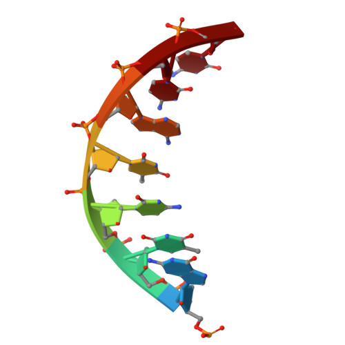
8XXK | pdb_00008xxk

Crystal structure of human 8-oxoguanine glycosylase K249H mutant bound to the reaction intermediate derived from the crystal soaked into the solution at pH 4.0 under 298 K for 3 weeks


Experimental Data Snapshot

  • Method: X-RAY DIFFRACTION
  • Resolution: 1.70 Å
  • R-Value Free: 
    0.213 (Depositor), 0.212 (DCC) 
  • R-Value Work: 
    0.163 (Depositor), 0.163 (DCC) 
  • R-Value Observed: 
    0.166 (Depositor) 

Starting Model: experimental
View more details

wwPDB Validation   3D Report Full Report


Ligand Structure Quality Assessment 


This is version 1.1 of the entry. See complete history


Literature

Capturing a glycosylase reaction intermediate in DNA repair by freeze-trapping of a pH-responsive hOGG1 mutant.

Unno, M.Morikawa, M.Sychrovsky, V.Koga, M.Minowa, N.Komuro, S.Shimizu, M.Fukuta, M.Tsuyuguchi, F.Mano, H.Ochi, Y.Nakashima, K.Okamoto, Y.Saio, T.Hattori, Y.Tanaka, Y.

(2025) Nucleic Acids Res 53

  • DOI: https://doi.org/10.1093/nar/gkaf718
  • Primary Citation of Related Structures:  
    8XWC, 8XWU, 8XXG, 8XXK

  • PubMed Abstract: 

    The human 8-oxoguanine DNA glycosylase 1 (hOGG1) is a bifunctional DNA repair enzyme that possesses both glycosylase and AP-lyase activity. Its AP-lyase reaction mechanism had been revealed by crystallographic capturing of the intermediate adduct. However, no intermediate within the glycosylase reaction was reported to date and the relevant reaction mechanism thus remained unresolved. In this work, we studied the glycosylase reaction of hOGG1 by time-resolved crystallography and spectroscopic/enzymological analyses. To trigger the glycosylase reaction within a crystal, we created a pH-responsive mutant of hOGG1 in which lysine 249 (K249) has been replaced by histidine (H), and designated hOGG1(K249H). Using hOGG1(K249H), a reactive intermediate state of the hOGG1(K249H)-DNA complex was captured in crystal upon pH activation. An unprecedented, ribose-ring-opened hemiaminal structure at the 8-oxoguanine (oxoG) site was found. Based on the structure of the reaction intermediate and QM/MM (quantum mechanics/molecular mechanics) calculations, a glycosylase reaction pathway of hOGG1(K249H) was identified where the aspartic acid 268 (D268) acts as a proton donor to O4' of oxoG. Moreover, enzymologically derived pKa (4.5) of a catalytic residue indicated that the observed pKa can be attributed to the carboxy group of D268. Thus, a reaction mechanism of the glycosylase reaction by hOGG1(K249H) has been proposed.


  • Organizational Affiliation
    • Graduate School of Science and Engineering, Ibaraki University, Hitachi, Ibaraki 316-8511, Japan.

Macromolecules

Find similar proteins by:  (by identity cutoff)  |  3D Structure
Entity ID: 3
MoleculeChains Sequence LengthOrganismDetailsImage
N-glycosylase/DNA lyaseC [auth A]336Homo sapiensMutation(s): 1 
Gene Names: OGG1MMHMUTMOGH1
EC: 3.2.2 (PDB Primary Data), 4.2.99.18 (PDB Primary Data)
UniProt & NIH Common Fund Data Resources
Find proteins for O15527 (Homo sapiens)
Explore O15527 
Go to UniProtKB:  O15527
PHAROS:  O15527
GTEx:  ENSG00000114026 
Entity Groups  
Sequence Clusters30% Identity50% Identity70% Identity90% Identity95% Identity100% Identity
UniProt GroupO15527
Sequence Annotations
Expand
  • Reference Sequence

Find similar nucleic acids by:  Sequence   |   3D Structure  

Entity ID: 1
MoleculeChains LengthOrganismImage
DNA (5'-D(*AP*GP*CP*GP*TP*CP*CP*A)-3')A [auth C]8Homo sapiens
Sequence Annotations
Expand
  • Reference Sequence

Find similar nucleic acids by:  Sequence   |   3D Structure  

Entity ID: 2
MoleculeChains LengthOrganismImage
DNA (5'-D(*GP*GP*TP*AP*GP*AP*CP*CP*TP*GP*GP*AP*CP*GP*C)-3')B [auth D]16Homo sapiens
Sequence Annotations
Expand
  • Reference Sequence

Find similar nucleic acids by:  Sequence   |   3D Structure  

Entity ID: 4
MoleculeChains LengthOrganismImage
DNA (5'-D(P*GP*TP*CP*TP*AP*CP*C)-3')D [auth G]7Homo sapiens
Sequence Annotations
Expand
  • Reference Sequence
Small Molecules
Ligands 4 Unique
IDChains Name / Formula / InChI Key2D Diagram3D Interactions
A1LXK (Subject of Investigation/LOI)
Query on A1LXK

Download Ideal Coordinates CCD File 
E [auth C][(2~{R},3~{S},5~{S})-5-[2-azanyl-6,8-bis(oxidanylidene)-1,7-dihydropurin-9-yl]-2,3,5-tris(oxidanyl)pentyl] dihydrogen phosphate
C10 H16 N5 O9 P
RDUREGGLHYLIPW-LMVFSUKVSA-N
GOL
Query on GOL

Download Ideal Coordinates CCD File 
F [auth D],
G [auth A],
H [auth A],
I [auth A]
GLYCEROL
C3 H8 O3
PEDCQBHIVMGVHV-UHFFFAOYSA-N
MG
Query on MG

Download Ideal Coordinates CCD File 
J [auth A],
K [auth A]
MAGNESIUM ION
Mg
JLVVSXFLKOJNIY-UHFFFAOYSA-N
NA
Query on NA

Download Ideal Coordinates CCD File 
L [auth A]SODIUM ION
Na
FKNQFGJONOIPTF-UHFFFAOYSA-N
Experimental Data & Validation

Experimental Data

  • Method: X-RAY DIFFRACTION
  • Resolution: 1.70 Å
  • R-Value Free:  0.213 (Depositor), 0.212 (DCC) 
  • R-Value Work:  0.163 (Depositor), 0.163 (DCC) 
  • R-Value Observed: 0.166 (Depositor) 
Space Group: P 21 21 21
Unit Cell:
Length ( Å )Angle ( ˚ )
a = 61.852α = 90
b = 69.448β = 90
c = 88.884γ = 90
Software Package:
Software NamePurpose
PHENIXrefinement
XDSdata reduction
Aimlessdata scaling
MOLREPphasing

Structure Validation

View Full Validation Report


Ligand Structure Quality Assessment 


Entry History & Funding Information

Deposition Data


Funding OrganizationLocationGrant Number
Japan Society for the Promotion of Science (JSPS)Japan22K05320
Japan Society for the Promotion of Science (JSPS)Japan19K05705
Japan Society for the Promotion of Science (JSPS)Japan16K07261
Japan Society for the Promotion of Science (JSPS)Japan19K06507

Revision History  (Full details and data files)

  • Version 1.0: 2025-07-23
    Type: Initial release
  • Version 1.1: 2025-08-13
    Changes: Database references