9VD8 | pdb_00009vd8

Structure of a truncated loopA mutant from the human gut flora K. grimontii Apg in complex with glucose


Experimental Data Snapshot

  • Method: X-RAY DIFFRACTION
  • Resolution: 2.39 Å
  • R-Value Free: 
    0.236 (Depositor), 0.237 (DCC) 
  • R-Value Work: 
    0.191 (Depositor), 0.191 (DCC) 
  • R-Value Observed: 
    0.193 (Depositor) 

Starting Model: experimental
View more details

wwPDB Validation   3D Report Full Report


Ligand Structure Quality Assessment 


This is version 1.0 of the entry. See complete history


Literature

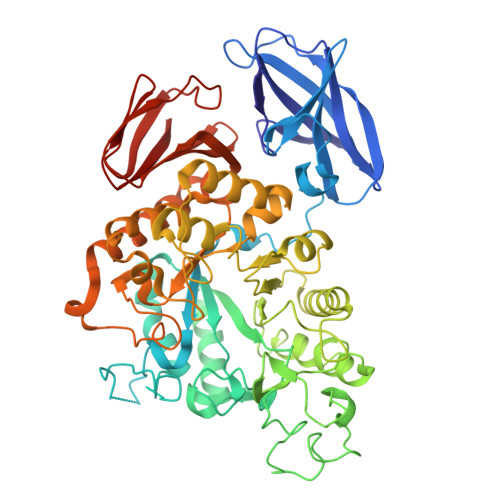
Molecular insights of acarbose metabolization catalyzed by acarbose-preferred glucosidase.

Huang, J.Shen, Z.Xiao, X.Wang, L.Zhang, J.Zhou, J.Gu, Y.

(2025) Nat Commun 16: 7839-7839

  • DOI: https://doi.org/10.1038/s41467-025-62855-y
  • Primary Citation of Related Structures:  
    9IVZ, 9IXH, 9IXW, 9IZE, 9IZO, 9UNS, 9V0I, 9VD8, 9VDG, 9VDW

  • PubMed Abstract: 

    The clinical efficacy of the antidiabetic drug acarbose is hampered by degradation by the acarbose-preferred glucosidase (Apg) from K. grimontii TD1. Understanding the catalytic mechanism of Apg can aid the design of next-generation hypoglycemic pharmaceuticals acarbose analogs. Here, we determine several crystal structures of Apg to identify the catalytic residues and the ligand-binding pocket of Apg. Structural analyses and computational modeling reveal D448 as the active nucleophile, contrasting with prior studies that assumed D336 to be the nucleophile. In addition to E373 proposed as the proton donor in previous reports, we find that R334 might be an alternative proton donor. Our experimental and computational analyses indicate the two-ring product acarviosine is the two-step hydrolyzed product, where the second hydrolysis is the rate-limiting step. Additionally, further investigation of the acarbose analogs acarstatins A and B that are resistant to Apg is conducted by computational analysis. Overall, our studies provide perspectives into the intricacies of Apg's catalytic mechanism, contributing to the design of next-generation hypoglycemic pharmaceuticals.


  • Organizational Affiliation
    • College of Chemistry & Pharmacy, Northwest A&F University, Yangling, Shaanxi, People's Republic of China.

Macromolecules
Find similar proteins by:  (by identity cutoff)  |  3D Structure
Entity ID: 1
MoleculeChains Sequence LengthOrganismDetailsImage
Apg-truncated loopA590Klebsiella grimontiiMutation(s): 0 
EC: 3.2.1.20
UniProt
Find proteins for A0ABD7ANA7 (Klebsiella grimontii)
Explore A0ABD7ANA7 
Go to UniProtKB:  A0ABD7ANA7
Entity Groups  
Sequence Clusters30% Identity50% Identity70% Identity90% Identity95% Identity100% Identity
UniProt GroupA0ABD7ANA7
Sequence Annotations
Expand
  • Reference Sequence
Small Molecules
Ligands 1 Unique
IDChains Name / Formula / InChI Key2D Diagram3D Interactions
GLC (Subject of Investigation/LOI)
Query on GLC

Download Ideal Coordinates CCD File 
B [auth A]alpha-D-glucopyranose
C6 H12 O6
WQZGKKKJIJFFOK-DVKNGEFBSA-N
Experimental Data & Validation

Experimental Data

  • Method: X-RAY DIFFRACTION
  • Resolution: 2.39 Å
  • R-Value Free:  0.236 (Depositor), 0.237 (DCC) 
  • R-Value Work:  0.191 (Depositor), 0.191 (DCC) 
  • R-Value Observed: 0.193 (Depositor) 
Space Group: P 65 2 2
Unit Cell:
Length ( Å )Angle ( ˚ )
a = 75.87α = 90
b = 75.87β = 90
c = 401.959γ = 120
Software Package:
Software NamePurpose
PHENIXrefinement
XDSdata reduction
Aimlessdata scaling
PHENIXphasing

Structure Validation

View Full Validation Report


Ligand Structure Quality Assessment 


Entry History & Funding Information

Deposition Data


Funding OrganizationLocationGrant Number
Not funded--

Revision History  (Full details and data files)

  • Version 1.0: 2025-09-03
    Type: Initial release