9DMM | pdb_00009dmm

Crystal structure of human KRAS G12C covalently bound to Divarasib (GDC6036)


Experimental Data Snapshot

  • Method: X-RAY DIFFRACTION
  • Resolution: 1.90 Å
  • R-Value Free: 
    0.228 (Depositor), 0.228 (DCC) 
  • R-Value Work: 
    0.170 (Depositor), 0.170 (DCC) 
  • R-Value Observed: 
    0.173 (Depositor) 

Starting Model: experimental
View more details

wwPDB Validation   3D Report Full Report


Ligand Structure Quality Assessment 


This is version 1.1 of the entry. See complete history


Literature

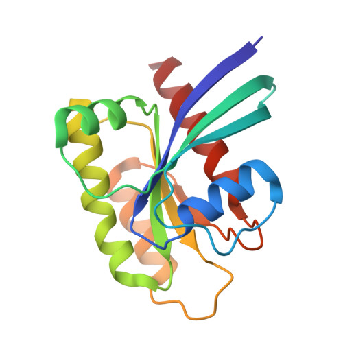
The structure of KRAS G12C bound to divarasib highlights features of potent switch-II pocket engagement.

Fernando, M.C.Craven, G.B.Shokat, K.M.

(2024) Small GTPases 15: 1-7

  • DOI: https://doi.org/10.1080/21541248.2025.2505441
  • Primary Citation of Related Structures:  
    9DMM

  • PubMed Abstract: 

    KRAS is the most frequently mutated oncogene in human cancer. In multiple types of cancer, a missense mutation at codon 12 substitutes a glycine for a cysteine, causing hyperactivation of the GTPase and enhanced MAPK signalling. Recent drug discovery efforts culminating from work during the past decade have resulted in two FDA-approved inhibitors, sotorasib and adagrasib, which target the KRAS G12C mutant allele. Ongoing medicinal chemistry efforts across academia and industry have continued developing more potent and efficacious KRAS G12C inhibitors. One agent in late-stage clinical trials, divarasib, has demonstrated robust overall response rates, in some cases greater than currently approved agents. Divarasib also exhibits enhanced covalent target engagement in vitro and significant specificity for KRAS G12C , yet the structural details of its binding have not been published. Here we report a high-resolution crystal structure of cysteine-light KRAS-4B G12C in complex with divarasib. Though it binds in the same allosteric pocket as sotorasib and adagrasib, the switch-II loop in each crystal structure takes on a distinct conformation differing as much as 5.6 Å between the Cα atom of residue 65 with sotorasib. Additionally, we highlight structural features of the drug complex that may guide future medicinal chemistry efforts targeting various KRAS alleles.


  • Organizational Affiliation
    • Department of Cellular and Molecular Pharmacology and Howard Hughes Medical Institute, University of California, San Francisco, CA, USA.

Macromolecules
Find similar proteins by:  (by identity cutoff)  |  3D Structure
Entity ID: 1
MoleculeChains Sequence LengthOrganismDetailsImage
Isoform 2B of GTPase KRas170Homo sapiensMutation(s): 4 
Gene Names: KRASKRAS2RASK2
EC: 3.6.5.2
UniProt & NIH Common Fund Data Resources
Find proteins for P01116 (Homo sapiens)
Explore P01116 
Go to UniProtKB:  P01116
PHAROS:  P01116
GTEx:  ENSG00000133703 
Entity Groups  
Sequence Clusters30% Identity50% Identity70% Identity90% Identity95% Identity100% Identity
UniProt GroupP01116
Sequence Annotations
Expand
  • Reference Sequence
Small Molecules
Ligands 3 Unique
IDChains Name / Formula / InChI Key2D Diagram3D Interactions
A1AWR (Subject of Investigation/LOI)
Query on A1AWR

Download Ideal Coordinates CCD File 
B [auth A]1-{(3S)-4-[(7M)-7-[6-amino-4-methyl-3-(trifluoromethyl)pyridin-2-yl]-6-chloro-8-fluoro-2-{[(2S)-1-methylpyrrolidin-2-yl]methoxy}quinazolin-4-yl]-3-methylpiperazin-1-yl}propan-1-one
C29 H34 Cl F4 N7 O2
QEAJZIRHKLXVLR-IRXDYDNUSA-N
GDP
Query on GDP

Download Ideal Coordinates CCD File 
D [auth A]GUANOSINE-5'-DIPHOSPHATE
C10 H15 N5 O11 P2
QGWNDRXFNXRZMB-UUOKFMHZSA-N
MG
Query on MG

Download Ideal Coordinates CCD File 
C [auth A]MAGNESIUM ION
Mg
JLVVSXFLKOJNIY-UHFFFAOYSA-N
Experimental Data & Validation

Experimental Data

  • Method: X-RAY DIFFRACTION
  • Resolution: 1.90 Å
  • R-Value Free:  0.228 (Depositor), 0.228 (DCC) 
  • R-Value Work:  0.170 (Depositor), 0.170 (DCC) 
  • R-Value Observed: 0.173 (Depositor) 
Space Group: P 21 21 21
Unit Cell:
Length ( Å )Angle ( ˚ )
a = 45.893α = 90
b = 59.626β = 90
c = 61.748γ = 90
Software Package:
Software NamePurpose
PHENIXrefinement
Aimlessdata scaling
XDSdata reduction
Cootmodel building
PHASERphasing

Structure Validation

View Full Validation Report


Ligand Structure Quality Assessment 


Entry History & Funding Information

Deposition Data


Funding OrganizationLocationGrant Number
National Institutes of Health/National Cancer Institute (NIH/NCI)United States5R01CA244550

Revision History  (Full details and data files)

  • Version 1.0: 2025-05-21
    Type: Initial release
  • Version 1.1: 2025-05-28
    Changes: Database references