9DP4 | pdb_00009dp4

APE1 N174Q Product Complex with Abasic DNA


Experimental Data Snapshot

  • Method: X-RAY DIFFRACTION
  • Resolution: 2.25 Å
  • R-Value Free: 
    0.248 (Depositor), 0.250 (DCC) 
  • R-Value Work: 
    0.215 (Depositor), 0.218 (DCC) 
  • R-Value Observed: 
    0.221 (Depositor) 

Starting Model: experimental
View more details

wwPDB Validation   3D Report Full Report


This is version 1.1 of the entry. See complete history


Literature

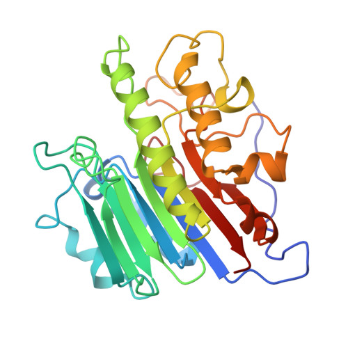
APE1 active site residue Asn174 stabilizes the AP site and is essential for catalysis.

DeHart, K.M.Hoitsma, N.M.Thompson, S.H.Borin, V.A.Agarwal, P.K.Freudenthal, B.D.

(2025) J Biological Chem 301: 110655-110655

  • DOI: https://doi.org/10.1016/j.jbc.2025.110655
  • Primary Citation of Related Structures:  
    9DP1, 9DP2, 9DP3, 9DP4

  • PubMed Abstract: 

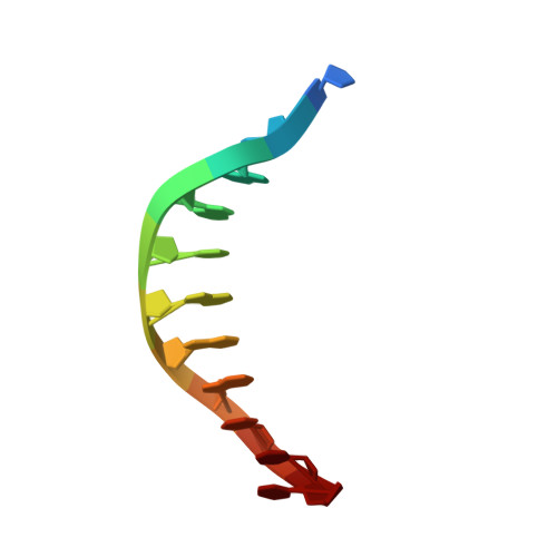
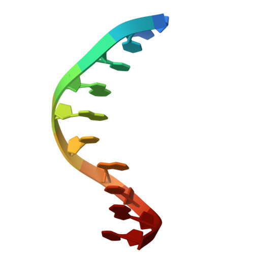
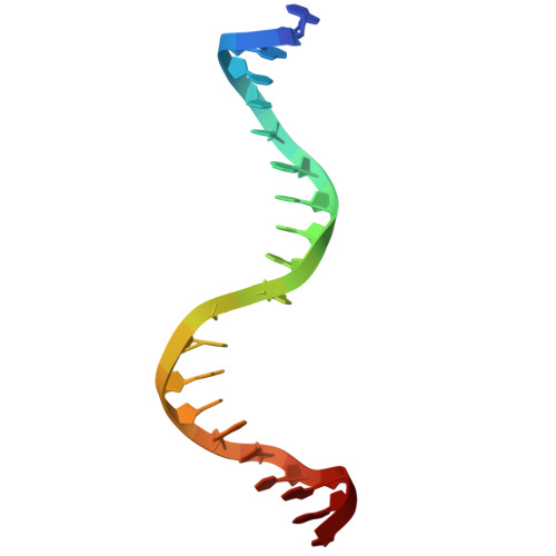
    Apurinic/apyrimidinic (AP) sites are common and highly mutagenic DNA lesions that can arise spontaneously or as intermediates during base excision repair. The enzyme apurinic/apyrimidinic endonuclease 1 (APE1) initiates repair of AP sites by cleaving the DNA backbone at the AP site via its endonuclease activity. Here, we investigated the functional role of the APE1 active site residue N174 that contacts the AP site during catalysis. To do so, we rationally designed four APE1 mutants (N174A, N174D, N174Q, and N174K) that altered the hydrogen bonding potential, electronegativity, and side chain length of residue 174. Kinetic analysis determined that AP site cleavage was impaired in our APE1 N174A , APE1 N174D , and APE1 N174K mutants, but not in our APE1 N174Q mutant. Using X-ray crystallography and molecular dynamic simulations of our APE1 mutants, we attributed the poor cleavage of our APE1 N174A and APE1 N174D mutants to lack of hydrogen bonding between residue 174 and the AP site. From these experiments, we also discovered that the altered charge of residue 174 in APE1 N174D and APE1 N174K mutants contributed to impaired cleavage. From analysis of our mutants, we determined that in APE1 WT , N174 stabilizes AP DNA within the APE1 active site via hydrogen bonding promoting cleavage of AP sites. We also found that the neutral charge of N174 in APE1 WT contributes to an optimal electrostatic environment during the APE1 endonuclease reaction resulting in efficient AP site cleavage. Cumulatively, we demonstrate the importance of N174 in APE1's function and provide new insights into the molecular mechanism by which APE1 processes AP sites during DNA repair.


  • Organizational Affiliation
    • Department of Biochemistry and Molecular Biology, University of Kansas Medical Center, Kansas City, Kansas, USA.

Macromolecules

Find similar proteins by:  (by identity cutoff)  |  3D Structure
Entity ID: 1
MoleculeChains Sequence LengthOrganismDetailsImage
DNA repair nuclease/redox regulator APEX1
A, B
276Homo sapiensMutation(s): 2 
Gene Names: APEX1APEAPE1APEXAPXHAP1REF1
EC: 3.1.11.2 (PDB Primary Data), 3.1.21 (PDB Primary Data)
UniProt & NIH Common Fund Data Resources
Find proteins for P27695 (Homo sapiens)
Explore P27695 
Go to UniProtKB:  P27695
PHAROS:  P27695
GTEx:  ENSG00000100823 
Entity Groups  
Sequence Clusters30% Identity50% Identity70% Identity90% Identity95% Identity100% Identity
UniProt GroupP27695
Sequence Annotations
Expand
  • Reference Sequence

Find similar nucleic acids by:  Sequence   |   3D Structure  

Entity ID: 2
MoleculeChains LengthOrganismImage
DNA (5'-D(P*(3DR)P*CP*GP*AP*CP*GP*GP*AP*TP*CP*C)-3')C [auth D],
F [auth G]
11synthetic construct
Sequence Annotations
Expand
  • Reference Sequence

Find similar nucleic acids by:  Sequence   |   3D Structure  

Entity ID: 3
MoleculeChains LengthOrganismImage
DNA (5'-D(*GP*CP*TP*GP*AP*TP*GP*CP*GP*C)-3')D [auth E],
G [auth H]
10synthetic construct
Sequence Annotations
Expand
  • Reference Sequence

Find similar nucleic acids by:  Sequence   |   3D Structure  

Entity ID: 4
MoleculeChains LengthOrganismImage
DNA (5'-D(*GP*GP*AP*TP*CP*CP*GP*TP*CP*GP*GP*GP*CP*GP*CP*AP*TP*CP*AP*GP*C)-3')E [auth F],
H [auth I]
21synthetic construct
Sequence Annotations
Expand
  • Reference Sequence
Experimental Data & Validation

Experimental Data

  • Method: X-RAY DIFFRACTION
  • Resolution: 2.25 Å
  • R-Value Free:  0.248 (Depositor), 0.250 (DCC) 
  • R-Value Work:  0.215 (Depositor), 0.218 (DCC) 
  • R-Value Observed: 0.221 (Depositor) 
Space Group: P 43
Unit Cell:
Length ( Å )Angle ( ˚ )
a = 153.423α = 90
b = 153.423β = 90
c = 45.373γ = 90
Software Package:
Software NamePurpose
PHENIXrefinement
HKL-3000data collection
HKL-3000data reduction
HKL-3000data scaling
PHENIXphasing
Cootmodel building
PDB_EXTRACTdata extraction

Structure Validation

View Full Validation Report


Entry History & Funding Information

Deposition Data


Funding OrganizationLocationGrant Number
National Institutes of Health/National Institute of Environmental Health Sciences (NIH/NIEHS)United StatesR01ES029203

Revision History  (Full details and data files)

  • Version 1.0: 2024-10-09
    Type: Initial release
  • Version 1.1: 2025-10-29
    Changes: Database references