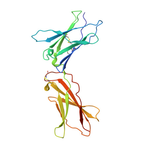
Design of a potent interleukin-21 mimic for cancer immunotherapy.
Chun, J.H., Lim, B.S., Roy, S., Walsh, M.J., Abhiraman, G.C., Zhangxu, K., Atajanova, T., Revach, O.Y., Clark, E.C., Li, P., Palin, C.A., Khanna, A., Tower, S., Kureshi, R., Hoffman, M.T., Sharova, T., Lawless, A., Cohen, S., Boland, G.M., Nguyen, T., Peprah, F., Tello, J.G., Liu, S.Y., Kim, C.J., Shin, H., Quijano-Rubio, A., Jude, K.M., Gerben, S., Murray, A., Heine, P., DeWitt, M., Ulge, U.Y., Carter, L., King, N.P., Silva, D.A., Kueh, H.Y., Kalia, V., Sarkar, S., Jenkins, R.W., Garcia, K.C., Leonard, W.J., Dougan, M., Dougan, S.K., Baker, D.(2025) Sci Immunol 10: eadx1582-eadx1582
- PubMed: 41004565
- DOI: https://doi.org/10.1126/sciimmunol.adx1582
- Primary Citation of Related Structures:
9E2T - PubMed Abstract:
Long-standing goals of cancer immunotherapy are to activate cytotoxic antitumor T cells across a range of affinities for tumor antigens while suppressing regulatory T cells. Computational protein design has enabled the precise tailoring of proteins to meet specific needs. Here, we report a de novo designed IL-21 mimic, 21h10, with high stability and signaling potency in humans and mice. In murine and ex vivo human organotypic tumor models, 21h10 showed robust antitumor activity, with more prolonged signaling and stronger antitumor activity than native IL-21. 21h10 induced pancreatitis that could be mitigated by TNF blockade without compromising antitumor efficacy. Although antidrug antibodies to 21h10 formed, they were not neutralizing. 21h10 induced highly cytotoxic T cells with a range of affinities, robustly expanding intratumoral low-affinity cytotoxic T cells and driving high expression of IFN-γ and granzyme B compared with native IL-21, while increasing the frequency of IFN-γ + T helper 1 cells and reducing regulatory T cells. The full human-mouse cross-reactivity, high stability and potency, and low-affinity antitumor responses support the translational potential of 21h10.
- Institute for Protein Design, University of Washington, Seattle, WA, USA.
Organizational Affiliation:


