9EG9 | pdb_00009eg9

Crystal structure of human dihydroorotate dehydrogenase in complex with lapachol


Experimental Data Snapshot

  • Method: X-RAY DIFFRACTION
  • Resolution: 1.31 Å
  • R-Value Free: 
    0.180 (Depositor), 0.190 (DCC) 
  • R-Value Work: 
    0.174 (Depositor), 0.183 (DCC) 
  • R-Value Observed: 
    0.174 (Depositor) 

Starting Model: experimental
View more details

wwPDB Validation   3D Report Full Report


Ligand Structure Quality Assessment 


This is version 1.0 of the entry. See complete history


Literature

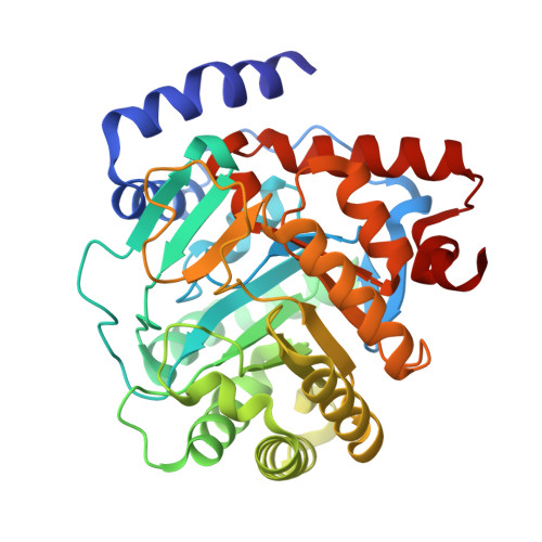
Crystallographic Structure of Human Dihydroorotate Dehydrogenase in Complex with the Natural Product Inhibitor Lapachol.

Purificacao, A.D.Benz, L.S.Lima Silva, W.J.Emery, F.S.Andrade, C.H.Weiss, M.S.Nonato, M.C.

(2025) ACS Omega 10: 29087-29097

  • DOI: https://doi.org/10.1021/acsomega.5c01536
  • Primary Citation of Related Structures:  
    9EG9

  • PubMed Abstract: 

    Dihydroorotate dehydrogenase (DHODH) is a key enzyme in the pyrimidine biosynthesis pathway, playing a critical role in cellular processes and offering therapeutic potential for antiviral, antineoplastic, and autoimmune treatments. Human DHODH ( Hs DHODH) utilizes ubiquinone as a second substrate, positioning its quinone-binding site as a promising target for inhibitor development. Lapachol, a natural naphthoquinone, has gained prominence as a valuable natural product for the discovery of novel therapeutic agents, thanks to its wide range of biological activities. In this study, we present the first crystal structure of Hs DHODH in complex with lapachol, providing valuable insights into the interactions between this natural product and the enzyme. The structure reveals key binding interactions that mediate lapachol's affinity for Hs DHODH and validates previously proposed computational models. Complementary molecular dynamics simulations further highlight the stability of the complex and the importance of water-mediated interactions in ligand binding. These findings enhance our understanding of how naphthoquinone derivatives, such as lapachol, interact with class 2 DHODHs, offering a foundation for the design of optimized inhibitors for therapeutic applications. By integration of structural and computational data, this study contributes to the rational design of novel Hs DHODH inhibitors, paving the way for future exploration of lapachol and its derivatives in drug discovery.


  • Organizational Affiliation

    Center for the Research and Advancement in Fragments and Molecular Targets (CRAFT), School of Pharmaceutical Sciences at Ribeirao Preto, University of São Paulo, Ribeirão Preto 14040-903, São Paulo, Brazil.


Macromolecules
Find similar proteins by:  (by identity cutoff)  |  3D Structure
Entity ID: 1
MoleculeChains Sequence LengthOrganismDetailsImage
Dihydroorotate dehydrogenase (quinone), mitochondrial368Homo sapiensMutation(s): 0 
Gene Names: DHODH
EC: 1.3.5.2
UniProt & NIH Common Fund Data Resources
Find proteins for Q02127 (Homo sapiens)
Explore Q02127 
Go to UniProtKB:  Q02127
PHAROS:  Q02127
GTEx:  ENSG00000102967 
Entity Groups  
Sequence Clusters30% Identity50% Identity70% Identity90% Identity95% Identity100% Identity
UniProt GroupQ02127
Sequence Annotations
Expand
  • Reference Sequence
Small Molecules
Ligands 6 Unique
IDChains Name / Formula / InChI Key2D Diagram3D Interactions
FMN (Subject of Investigation/LOI)
Query on FMN

Download Ideal Coordinates CCD File 
B [auth A]FLAVIN MONONUCLEOTIDE
C17 H21 N4 O9 P
FVTCRASFADXXNN-SCRDCRAPSA-N
A1BIJ (Subject of Investigation/LOI)
Query on A1BIJ

Download Ideal Coordinates CCD File 
I [auth A]2-hydroxy-3-(3-methylbut-2-en-1-yl)naphthalene-1,4-dione
C15 H14 O3
CIEYTVIYYGTCCI-UHFFFAOYSA-N
ORO (Subject of Investigation/LOI)
Query on ORO

Download Ideal Coordinates CCD File 
C [auth A]OROTIC ACID
C5 H4 N2 O4
PXQPEWDEAKTCGB-UHFFFAOYSA-N
SO4
Query on SO4

Download Ideal Coordinates CCD File 
D [auth A],
E [auth A],
G [auth A]
SULFATE ION
O4 S
QAOWNCQODCNURD-UHFFFAOYSA-L
GOL
Query on GOL

Download Ideal Coordinates CCD File 
H [auth A],
J [auth A],
K [auth A]
GLYCEROL
C3 H8 O3
PEDCQBHIVMGVHV-UHFFFAOYSA-N
ACT
Query on ACT

Download Ideal Coordinates CCD File 
F [auth A]ACETATE ION
C2 H3 O2
QTBSBXVTEAMEQO-UHFFFAOYSA-M
Experimental Data & Validation

Experimental Data

  • Method: X-RAY DIFFRACTION
  • Resolution: 1.31 Å
  • R-Value Free:  0.180 (Depositor), 0.190 (DCC) 
  • R-Value Work:  0.174 (Depositor), 0.183 (DCC) 
  • R-Value Observed: 0.174 (Depositor) 
Space Group: P 32 2 1
Unit Cell:
Length ( Å )Angle ( ˚ )
a = 90.46α = 90
b = 90.46β = 90
c = 122.66γ = 120
Software Package:
Software NamePurpose
REFMACrefinement
pointlessdata scaling
XDSdata reduction
PHASERphasing

Structure Validation

View Full Validation Report


Ligand Structure Quality Assessment 


Entry History & Funding Information

Deposition Data


Funding OrganizationLocationGrant Number
Brazilian National Council for Scientific and Technological Development (CNPq)Brazil441038/2020-4
Sao Paulo Research Foundation (FAPESP)Brazil2021/13237-5
Sao Paulo Research Foundation (FAPESP)Brazil2020/06190-0
iNEXT-DiscoveryEuropean Union871037
German Research Foundation (DFG)GermanyFE2166/1-1

Revision History  (Full details and data files)

  • Version 1.0: 2025-08-06
    Type: Initial release