Structure of the Pseudomonas aeruginosa PAO1 Type IV pilus.
Ochner, H., Bohning, J., Wang, Z., Tarafder, A.K., Caspy, I., Bharat, T.A.M.(2024) PLoS Pathog 20: e1012773-e1012773
- PubMed: 39666767
- DOI: https://doi.org/10.1371/journal.ppat.1012773
- Primary Citation of Related Structures:
9EWX - PubMed Abstract:
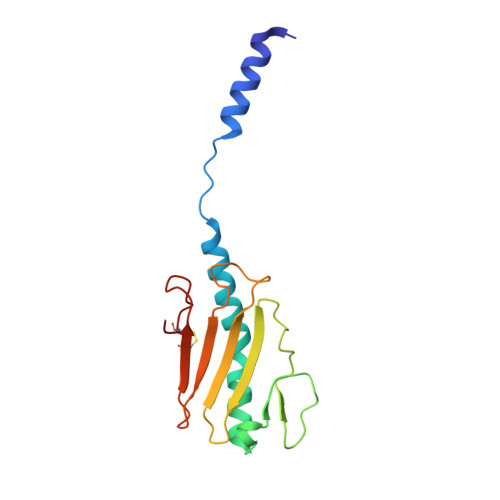
Type IV pili (T4Ps) are abundant in many bacterial and archaeal species, where they play important roles in both surface sensing and twitching motility, with implications for adhesion, biofilm formation and pathogenicity. While Type IV pilus (T4P) structures from other organisms have been previously solved, a high-resolution structure of the native, fully assembled T4P of Pseudomonas aeruginosa, a major human pathogen, would be valuable in a drug discovery context. Here, we report a 3.2 Å-resolution structure of the P. aeruginosa PAO1 T4P determined by electron cryomicroscopy (cryo-EM). PilA subunits constituting the T4P exhibit a classical pilin fold featuring an extended N-terminal α-helix linked to a C-terminal globular β-sheet-containing domain, which are packed tightly along the pilus, in line with models derived from previous cryo-EM data of the P. aeruginosa PAK strain. The N-terminal helices constitute the pilus core where they stabilise the tubular assembly via hydrophobic interactions. The α-helical core of the pilus is surrounded by the C-terminal globular domain of PilA that coats the outer surface of the pilus, mediating interactions with the surrounding environment. Comparison of the P. aeruginosa PAO1 T4P with T4P structures from other organisms, both at the level of the pilin subunits and the fully assembled pili, confirms previously described common architectural principles whilst highlighting key differences between members of this abundant class of prokaryotic filaments. This study provides a structural framework for understanding the molecular and cell biology of these important cellular appendages mediating interaction of prokaryotes to surfaces.
- Structural Studies Division, MRC Laboratory of Molecular Biology, Francis Crick Avenue, Cambridge, United Kingdom.
Organizational Affiliation: