


Crystal structure of human Dihydroorotate Dehydrogenase in complex with the inhibitor N-(3'-(2,2-difluoropropoxy)-2,3,5,6-tetrafluoro-[1,1'-biphenyl]-4-yl)-2-hydroxypyrazolo[1,5-a]pyridine-3-carboxamide
Alberti, M., Miggiano, R.To be published.
Experimental Data Snapshot
Starting Model: experimental
View more details
Entity ID: 1 | |||||
|---|---|---|---|---|---|
| Molecule | Chains | Sequence Length | Organism | Details | Image |
| Dihydroorotate dehydrogenase (quinone), mitochondrial | 390 | Homo sapiens | Mutation(s): 0 Gene Names: DHODH EC: 1.3.5.2 | 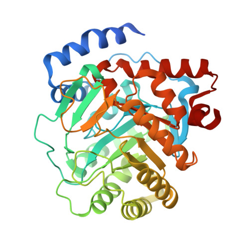 | |
UniProt & NIH Common Fund Data Resources | |||||
Find proteins for Q02127 (Homo sapiens) Explore Q02127 Go to UniProtKB: Q02127 | |||||
PHAROS: Q02127 GTEx: ENSG00000102967 | |||||
Entity Groups | |||||
| Sequence Clusters | 30% Identity50% Identity70% Identity90% Identity95% Identity100% Identity | ||||
| UniProt Group | Q02127 | ||||
Sequence AnnotationsExpand | |||||
| |||||
| Ligands 5 Unique | |||||
|---|---|---|---|---|---|
| ID | Chains | Name / Formula / InChI Key | 2D Diagram | 3D Interactions | |
| A1H8I (Subject of Investigation/LOI) Query on A1H8I | E [auth A] | N-[4-[3-[2,2-bis(fluoranyl)propoxy]phenyl]-2,3,5,6-tetrakis(fluoranyl)phenyl]-2-oxidanyl-pyrazolo[1,5-a]pyridine-3-carboxamide C23 H15 F6 N3 O3 VMNSFPULVHCEOI-UHFFFAOYSA-N |  | ||
| FMN Query on FMN | C [auth A] | FLAVIN MONONUCLEOTIDE C17 H21 N4 O9 P FVTCRASFADXXNN-SCRDCRAPSA-N |  | ||
| P6G Query on P6G | D [auth A] | HEXAETHYLENE GLYCOL C12 H26 O7 IIRDTKBZINWQAW-UHFFFAOYSA-N |  | ||
| ORO Query on ORO | B [auth A] | OROTIC ACID C5 H4 N2 O4 PXQPEWDEAKTCGB-UHFFFAOYSA-N |  | ||
| SO4 Query on SO4 | F [auth A] | SULFATE ION O4 S QAOWNCQODCNURD-UHFFFAOYSA-L |  | ||
| Length ( Å ) | Angle ( ˚ ) |
|---|---|
| a = 91.071 | α = 90 |
| b = 91.071 | β = 90 |
| c = 123.441 | γ = 120 |
| Software Name | Purpose |
|---|---|
| PHENIX | refinement |
| SCALA | data scaling |
| XDS | data reduction |
| PHASER | phasing |
| Funding Organization | Location | Grant Number |
|---|---|---|
| Ministero dell Universita e della Ricerca | Italy | -- |


