Cryo-EM studies of amyloid-beta fibrils from human and murine brains carrying the Uppsala APP mutation ( Delta 690-695).
Zielinski, M., Peralta Reyes, F.S., Gremer, L., Sommerhage, S., Pagnon de la Vega, M., Roder, C., Heidler, T.V., Syvanen, S., Willbold, D., Sehlin, D., Ingelsson, M., Schroder, G.F.(2025) Acta Neuropathol Commun 13: 209-209
- PubMed: 41044681
- DOI: https://doi.org/10.1186/s40478-025-02120-x
- Primary Citation of Related Structures:
9FH1, 9FH2, 9FH3, 9FH4, 9FH5, 9FH6 - PubMed Abstract:
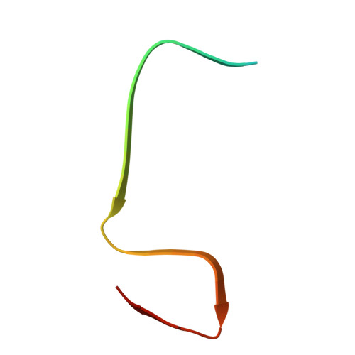
Today, 13 intra-amyloid-β (Aβ) amyloid precursor protein (APP) gene mutations are known to cause familial Alzheimer's disease (AD). Most of them are point mutations causing an increased production or a change in the conformation of Aβ. The Uppsala APP mutation (Δ690-695 in APP, Δ19-24 in Aβ) is the first known multi-codon deletion causing autosomal dominant AD. Here, we applied cryo-electron microscopy (cryo-EM) to investigate the structure of Aβ fibrils with the Uppsala APP mutation from tg-UppSwe mouse brain tissue. Murine AβUpp(1-42) Δ19-24 are made of two identical S-shaped protofilaments with an ordered fibril core of S8-A42. The murine Aβ fold is almost identical to previously described human type II filaments, although the amino acid sequences differ considerably. In addition, we report the cryo-EM structure of Aβ fibrils from the temporal cortex of a patient with the Uppsala APP mutation. The observed structure of the human Aβ fold closely resembles previously described type I fibrils. Structural modeling suggests that these fibrils are composed of wild-type Aβ, which implies that AβUpp may be less soluble and thus not readily accessible for cryo-EM image processing and structure determination. Additionally, from the human sample we determined the structures of tau paired helical filaments and tau straight filaments, which are identical to those found in sporadic AD cases. Finally, we present the 3D cryo-EM structures of four dominant AβUpp(1-42) Δ19-24 fibril polymorphs, formed in vitro. All four polymorphs differ from the observed folds of Uppsala Aβ in murine and human brain tissue, respectively.
- Institute of Biological Information Processing, Structural Biochemistry (IBI-7), Forschungszentrum Jülich, Jülich, Germany.
Organizational Affiliation: