9FQC | pdb_00009fqc

Crystal structure of phosphoserine phosphatase (SerB) from Brucella melitensis in copmplex with O-Phosphoserine


Experimental Data Snapshot

  • Method: X-RAY DIFFRACTION
  • Resolution: 2.35 Å
  • R-Value Free: 
    0.193 (Depositor), 0.194 (DCC) 
  • R-Value Work: 
    0.160 (Depositor), 0.162 (DCC) 
  • R-Value Observed: 
    0.161 (Depositor) 

Starting Model: experimental
View more details

wwPDB Validation   3D Report Full Report


Ligand Structure Quality Assessment 


This is version 1.1 of the entry. See complete history


Literature

Structural and Enzymological Characterization of Phosphoserine Phosphatase From Brucella melitensis.

Scaillet, T.Pierson, E.Fillet, M.Wouters, J.

(2025) Proteins 

  • DOI: https://doi.org/10.1002/prot.70027
  • Primary Citation of Related Structures:  
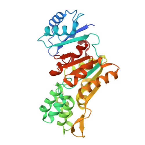
    8Q4S, 8QOB, 9FQ5, 9FQC, 9FQN

  • PubMed Abstract: 

    Amino acid L-serine (L-Ser) is a precursor of various biomolecules, including other amino acids, glutathione, and nucleotides. The metabolism of this amino acid is crucial in diseases such as brucellosis. Previous studies have revealed that the enzymes involved in L-Ser biosynthesis are essential for Brucella replication, making them potential targets for the development of new drugs. Here, we focus on Brucella melitensis phosphoserine phosphatase (BmPSP), which catalyzes the dephosphorylation of phosphoserine in L-Ser. The enzyme is characterized through enzymatic and structural studies, leading to the discovery of its first crystallographic structures. The interactions of BmPSP with different ligands are also investigated. We demonstrate that the substitution of its Mg 2+ cofactor with Ca 2+ inhibits the enzyme and results in a slight movement of catalytic residues in the active site. Crystallographic structures of BmPSP in complex with substrate, reaction products, and substrate analogs are also detailed, revealing the interaction between these molecules and the active site residues. This structural study provides a better understanding of phosphoserine phosphatases, highlighting the involvement of two highly conserved residues in the mechanism of substrate entry into the active site.


  • Organizational Affiliation

    Department of Chemistry, Laboratoire de Chimie Biologique Structurale (CBS), University of Namur (UNamur), Namur Research Institute for Life Sciences (NARILIS), Namur Institute of Structural Matter (NISM), Namur, Belgium.


Macromolecules
Find similar proteins by:  (by identity cutoff)  |  3D Structure
Entity ID: 1
MoleculeChains Sequence LengthOrganismDetailsImage
Phosphoserine phosphatase307Brucella melitensisMutation(s): 0 
Gene Names: BMEI0615
EC: 3.1.3.3
UniProt
Find proteins for Q8YI30 (Brucella melitensis biotype 1 (strain ATCC 23456 / CCUG 17765 / NCTC 10094 / 16M))
Explore Q8YI30 
Go to UniProtKB:  Q8YI30
Entity Groups  
Sequence Clusters30% Identity50% Identity70% Identity90% Identity95% Identity100% Identity
UniProt GroupQ8YI30
Sequence Annotations
Expand
  • Reference Sequence
Small Molecules
Experimental Data & Validation

Experimental Data

  • Method: X-RAY DIFFRACTION
  • Resolution: 2.35 Å
  • R-Value Free:  0.193 (Depositor), 0.194 (DCC) 
  • R-Value Work:  0.160 (Depositor), 0.162 (DCC) 
  • R-Value Observed: 0.161 (Depositor) 
Space Group: I 21 3
Unit Cell:
Length ( Å )Angle ( ˚ )
a = 145.25α = 90
b = 145.25β = 90
c = 145.25γ = 90
Software Package:
Software NamePurpose
MxCuBEdata collection
PHENIXrefinement
XDSdata reduction
XSCALEdata scaling
PHASERphasing
Cootmodel building

Structure Validation

View Full Validation Report


Ligand Structure Quality Assessment 


Entry History & Funding Information

Deposition Data


Funding OrganizationLocationGrant Number
Not funded--

Revision History  (Full details and data files)

  • Version 1.0: 2025-06-25
    Type: Initial release
  • Version 1.1: 2025-08-06
    Changes: Database references