9G2U | pdb_00009g2u

Endophilin B1 dimer bound to nanodisc center


Experimental Data Snapshot

  • Method: ELECTRON MICROSCOPY
  • Resolution: 3.45 Å
  • Aggregation State: PARTICLE 
  • Reconstruction Method: SINGLE PARTICLE 

Starting Model: in silico
View more details

wwPDB Validation   3D Report Full Report


This is version 1.2 of the entry. See complete history


Literature

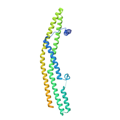
Peripheral membrane protein endophilin B1 probes, perturbs and permeabilizes lipid bilayers.

Thorlacius, A.Rulev, M.Sundberg, O.Sundborger-Lunna, A.

(2025) Commun Biol 8: 182-182

  • DOI: https://doi.org/10.1038/s42003-025-07610-1
  • Primary Citation of Related Structures:  
    9G2R, 9G2U, 9G2W

  • PubMed Abstract: 

    Bin/Amphiphysin/Rvs167 (BAR) domain containing proteins are peripheral membrane proteins that regulate intracellular membrane curvature. BAR protein endophilin B1 plays a key role in multiple cellular processes critical for oncogenesis, including autophagy and apoptosis. Amphipathic regions in endophilin B1 drive membrane association and tubulation through membrane scaffolding. Our understanding of exactly how BAR proteins like endophilin B1 promote highly diverse intracellular membrane remodeling events in the cell is severely limited due to lack of high-resolution structural information. Here we present the highest resolution cryo-EM structure of a BAR protein to date and the first structures of a BAR protein bound to a lipid bicelle. Using neural networks, we can effectively sort particle species of different stoichiometries, revealing the tremendous flexibility of post-membrane binding, pre-polymer BAR dimer organization and membrane deformation. We also show that endophilin B1 efficiently permeabilizes negatively charged liposomes that contain mitochondria-specific lipid cardiolipin and propose a new model for Bax-mediated cell death.


  • Organizational Affiliation
    • Department of Cell and Molecular Biology, Uppsala University, Uppsala, Sweden.

Macromolecules
Find similar proteins by:  (by identity cutoff)  |  3D Structure
Entity ID: 1
MoleculeChains Sequence LengthOrganismDetailsImage
Endophilin-B1
A, B
365Homo sapiensMutation(s): 0 
Gene Names: SH3GLB1KIAA0491CGI-61
UniProt & NIH Common Fund Data Resources
Find proteins for Q9Y371 (Homo sapiens)
Explore Q9Y371 
Go to UniProtKB:  Q9Y371
PHAROS:  Q9Y371
GTEx:  ENSG00000097033 
Entity Groups  
Sequence Clusters30% Identity50% Identity70% Identity90% Identity95% Identity100% Identity
UniProt GroupQ9Y371
Sequence Annotations
Expand
  • Reference Sequence
Experimental Data & Validation

Experimental Data

  • Method: ELECTRON MICROSCOPY
  • Resolution: 3.45 Å
  • Aggregation State: PARTICLE 
  • Reconstruction Method: SINGLE PARTICLE 
EM Software:
TaskSoftware PackageVersion
RECONSTRUCTIONcryoSPARC
MODEL REFINEMENTServalcat
MODEL REFINEMENTPHENIX
MODEL REFINEMENTCoot

Structure Validation

View Full Validation Report


Entry History & Funding Information

Deposition Data


Funding OrganizationLocationGrant Number
Swedish Research CouncilSweden2021-05423

Revision History  (Full details and data files)

  • Version 1.0: 2024-08-07
    Type: Initial release
  • Version 1.1: 2024-09-04
    Changes: Data collection, Database references
  • Version 1.2: 2025-02-19
    Changes: Data collection, Database references, Structure summary