9H2X | pdb_00009h2x

Crystal structure of stabilized A2A adenosine receptor A2AR-StaR2-bRIL in complex with compound 7, a novel nanomolar A2A receptor antagonist from modern hit-finding with structure-guided de novo design


Experimental Data Snapshot

  • Method: X-RAY DIFFRACTION
  • Resolution: 1.75 Å
  • R-Value Free: 
    0.226 (Depositor), 0.222 (DCC) 
  • R-Value Work: 
    0.206 (Depositor), 0.201 (DCC) 
  • R-Value Observed: 
    0.207 (Depositor) 

Starting Model: experimental
View more details

wwPDB Validation   3D Report Full Report


Ligand Structure Quality Assessment 


This is version 1.1 of the entry. See complete history


Literature

Identification of nanomolar adenosine A 2A receptor ligands using reinforcement learning and structure-based drug design.

Thomas, M.Matricon, P.G.Gillespie, R.J.Napiorkowska, M.Neale, H.Mason, J.S.Brown, J.Harwood, K.Fieldhouse, C.Swain, N.A.Geng, T.O'Boyle, N.M.Deflorian, F.Bender, A.de Graaf, C.

(2025) Nat Commun 16: 5485-5485

  • DOI: https://doi.org/10.1038/s41467-025-60629-0
  • Primary Citation of Related Structures:  
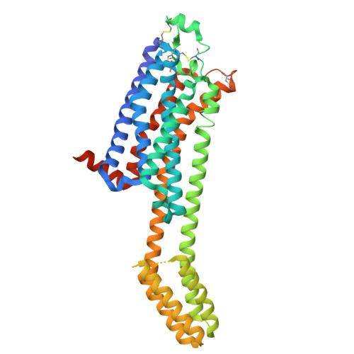
    9H2X, 9H37

  • PubMed Abstract: 

    Generative chemical language models (CLMs) have demonstrated success in learning language-based molecular representations for de novo drug design. Here, we integrate structure-based drug design (SBDD) principles with CLMs to go from protein structure to novel small-molecule ligands, without a priori knowledge of ligand chemistry. Using Augmented Hill-Climb, we successfully optimise multiple objectives within a practical timeframe, including protein-ligand complementarity. Resulting de novo molecules contain known or promising adenosine A 2A receptor ligand chemistry that is not available in commercial vendor libraries, accessing commercially novel areas of chemical space. Experimental validation demonstrates a binding hit rate of 88%, with 50% having confirmed functional activity, including three nanomolar ligands and two novel chemotypes. The two strongest binders are co-crystallised with the A 2A receptor, revealing their binding mechanisms that can be used to inform future iterations of structure-based de novo design, closing the AI SBDD loop.


  • Organizational Affiliation
    • Centre for Molecular Informatics, Department of Chemistry, University of Cambridge, Cambridge, UK.

Macromolecules
Find similar proteins by:  (by identity cutoff)  |  3D Structure
Entity ID: 1
MoleculeChains Sequence LengthOrganismDetailsImage
Adenosine receptor A2a,Soluble cytochrome b562433Homo sapiensEscherichia coliMutation(s): 0 
Gene Names: ADORA2AADORA2cybC
UniProt & NIH Common Fund Data Resources
Find proteins for P29274 (Homo sapiens)
Explore P29274 
Go to UniProtKB:  P29274
PHAROS:  P29274
GTEx:  ENSG00000128271 
Entity Groups  
Sequence Clusters30% Identity50% Identity70% Identity90% Identity95% Identity100% Identity
UniProt GroupP29274
Sequence Annotations
Expand
  • Reference Sequence
Small Molecules
Ligands 4 Unique
IDChains Name / Formula / InChI Key2D Diagram3D Interactions
CLR
Query on CLR

Download Ideal Coordinates CCD File 
C [auth A],
D [auth A],
E [auth A]
CHOLESTEROL
C27 H46 O
HVYWMOMLDIMFJA-DPAQBDIFSA-N
A1IR1 (Subject of Investigation/LOI)
Query on A1IR1

Download Ideal Coordinates CCD File 
AA [auth A]4-(furan-2-yl)-6-(6-imidazol-1-ylpyridin-2-yl)-1,3,5-triazin-2-amine
C15 H11 N7 O
PZPYYFUYDDSXJF-UHFFFAOYSA-N
OLA
Query on OLA

Download Ideal Coordinates CCD File 
F [auth A]
G [auth A]
H [auth A]
I [auth A]
J [auth A]
F [auth A],
G [auth A],
H [auth A],
I [auth A],
J [auth A],
K [auth A],
L [auth A],
M [auth A],
N [auth A],
O [auth A],
P [auth A],
Q [auth A],
R [auth A],
S [auth A],
T [auth A],
U [auth A],
V [auth A],
W [auth A],
X [auth A],
Y [auth A],
Z [auth A]
OLEIC ACID
C18 H34 O2
ZQPPMHVWECSIRJ-KTKRTIGZSA-N
NA
Query on NA

Download Ideal Coordinates CCD File 
B [auth A]SODIUM ION
Na
FKNQFGJONOIPTF-UHFFFAOYSA-N
Experimental Data & Validation

Experimental Data

  • Method: X-RAY DIFFRACTION
  • Resolution: 1.75 Å
  • R-Value Free:  0.226 (Depositor), 0.222 (DCC) 
  • R-Value Work:  0.206 (Depositor), 0.201 (DCC) 
  • R-Value Observed: 0.207 (Depositor) 
Space Group: C 2 2 21
Unit Cell:
Length ( Å )Angle ( ˚ )
a = 39.349α = 90
b = 179.535β = 90
c = 140.115γ = 90
Software Package:
Software NamePurpose
BUSTERrefinement
autoPROCdata processing
autoPROCdata reduction
Aimlessdata scaling
PHASERphasing

Structure Validation

View Full Validation Report


Ligand Structure Quality Assessment 


Entry History & Funding Information

Deposition Data


Funding OrganizationLocationGrant Number
University of CambridgeUnited Kingdom--

Revision History  (Full details and data files)

  • Version 1.0: 2025-06-18
    Type: Initial release
  • Version 1.1: 2025-07-16
    Changes: Database references