


Phosphorylation of Pannexin 1 activates ATP release
Zhang, Q., Gaullier, G., Dahl, G., Mim, C.To be published.
Experimental Data Snapshot
Starting Model: in silico
View more details
wwPDB Validation 3D Report Full Report

Entity ID: 1 | |||||
|---|---|---|---|---|---|
| Molecule | Chains | Sequence Length | Organism | Details | Image |
| Pannexin-1 | 444 | Mus musculus | Mutation(s): 0 Gene Names: Panx1 | 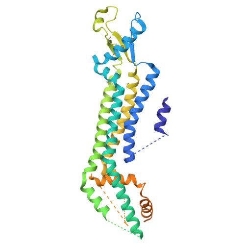 | |
UniProt & NIH Common Fund Data Resources | |||||
Find proteins for Q9JIP4 (Mus musculus) Explore Q9JIP4 Go to UniProtKB: Q9JIP4 | |||||
IMPC: MGI:1860055 | |||||
Entity Groups | |||||
| Sequence Clusters | 30% Identity50% Identity70% Identity90% Identity95% Identity100% Identity | ||||
| UniProt Group | Q9JIP4 | ||||
Sequence AnnotationsExpand | |||||
| |||||
| Task | Software Package | Version |
|---|---|---|
| MODEL REFINEMENT | PHENIX | 1.21.2_5419 |
| Funding Organization | Location | Grant Number |
|---|---|---|
| Carl Trygger Foundation | Sweden | CTS 21:1630 |


