9HMF | pdb_00009hmf

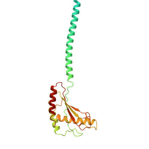
Periplasmic scaffold of the Campylobacter jejuni flagellar motor (alpha carbon trace)

  • Classification: STRUCTURAL PROTEIN
  • Organism(s): Campylobacter jejuni
  • Mutation(s): No 

  • Deposited: 2024-12-09 Released: 2024-12-25 
  • Deposition Author(s): Drobnic, T., Beeby, M.
  • Funding Organization(s): Biotechnology and Biological Sciences Research Council (BBSRC), Medical Research Council (MRC, United Kingdom), Human Frontier Science Program (HFSP), National Institutes of Health/National Institute Of Allergy and Infectious Diseases (NIH/NIAID), Cancer Research UK, Wellcome Trust

Experimental Data Snapshot

  • Method: ELECTRON MICROSCOPY
  • Resolution: 7.90 Å
  • Aggregation State: PARTICLE 
  • Reconstruction Method: SINGLE PARTICLE 

Starting Models: in silico
View more details

wwPDB Validation   3D Report Full Report


This is version 1.1 of the entry. See complete history


Literature

In situ structure of a bacterial flagellar motor at subnanometre resolution reveals adaptations for increased torque.

Drobnic, T.Cohen, E.J.Calcraft, T.Alzheimer, M.Froschauer, K.Svensson, S.Hoffman, W.H.Singh, N.Garg, S.G.Henderson, L.D.Umrekar, T.R.Nans, A.Ribardo, D.Pedaci, F.Nord, A.L.Hochberg, G.K.A.Hendrixson, D.R.Sharma, C.M.Rosenthal, P.B.Beeby, M.

(2025) Nat Microbiol 10: 1723-1740

  • DOI: https://doi.org/10.1038/s41564-025-02012-9
  • Primary Citation of Related Structures:  
    9HMF

  • PubMed Abstract: 

    The bacterial flagellar motor, which spins a helical propeller for propulsion, has undergone evolutionary diversification across bacterial species, often involving the addition of structures associated with increasing torque for motility in viscous environments. Understanding how such structures function and have evolved is hampered by challenges in visualizing motors in situ. Here we developed a Campylobacter jejuni minicell system for in situ cryogenic electron microscopy imaging and single-particle analysis of its motor, one of the most complex flagellar motors known, to subnanometre resolution. Focusing on the large periplasmic structures which are essential for increasing torque, our structural data, interpreted with molecular models, show that the basal disk comprises concentric rings of FlgP. The medial disk is a lattice of PflC with PflD, while the proximal disk is a rim of PflB attached to spokes of PflA. PflAB dimerization is essential for proximal disk assembly, recruiting FliL to scaffold more stator complexes at a wider radius which increases torque. We also acquired insights into universal principles of flagellar torque generation. This in situ approach is broadly applicable to other membrane-residing bacterial molecular machines.


  • Organizational Affiliation
    • Department of Life Sciences, Imperial College London, London, UK.

Macromolecules
Find similar proteins by:  (by identity cutoff)  |  3D Structure
Entity ID: 1
MoleculeChains Sequence LengthOrganismDetailsImage
Flagellar protein FliLA [auth O],
B [auth P],
C [auth Q],
D [auth R]
178Campylobacter jejuniMutation(s): 0 
UniProt
Find proteins for A0A0H3PIF6 (Campylobacter jejuni subsp. jejuni serotype O:23/36 (strain 81-176))
Explore A0A0H3PIF6 
Go to UniProtKB:  A0A0H3PIF6
Entity Groups  
Sequence Clusters30% Identity50% Identity70% Identity90% Identity95% Identity100% Identity
UniProt GroupA0A0H3PIF6
Sequence Annotations
Expand
  • Reference Sequence
Find similar proteins by:  (by identity cutoff)  |  3D Structure
Entity ID: 2
MoleculeChains Sequence LengthOrganismDetailsImage
Paralyzed flagella protein PflAE [auth K]788Campylobacter jejuniMutation(s): 0 
UniProt
Find proteins for A0A0H3PAU5 (Campylobacter jejuni subsp. jejuni serotype O:23/36 (strain 81-176))
Explore A0A0H3PAU5 
Go to UniProtKB:  A0A0H3PAU5
Entity Groups  
Sequence Clusters30% Identity50% Identity70% Identity90% Identity95% Identity100% Identity
UniProt GroupA0A0H3PAU5
Sequence Annotations
Expand
  • Reference Sequence
Find similar proteins by:  (by identity cutoff)  |  3D Structure
Entity ID: 3
MoleculeChains Sequence LengthOrganismDetailsImage
TPR domain proteinF [auth L]820Campylobacter jejuniMutation(s): 0 
UniProt
Find proteins for A0A0H3PJ87 (Campylobacter jejuni subsp. jejuni serotype O:23/36 (strain 81-176))
Explore A0A0H3PJ87 
Go to UniProtKB:  A0A0H3PJ87
Entity Groups  
Sequence Clusters30% Identity50% Identity70% Identity90% Identity95% Identity100% Identity
UniProt GroupA0A0H3PJ87
Sequence Annotations
Expand
  • Reference Sequence
Find similar proteins by:  (by identity cutoff)  |  3D Structure
Entity ID: 4
MoleculeChains Sequence LengthOrganismDetailsImage
PDZ domain-containing protein364Campylobacter jejuniMutation(s): 0 
UniProt
Find proteins for A0A1E7NUR8 (Campylobacter jejuni)
Explore A0A1E7NUR8 
Go to UniProtKB:  A0A1E7NUR8
Entity Groups  
Sequence Clusters30% Identity50% Identity70% Identity90% Identity95% Identity100% Identity
UniProt GroupA0A1E7NUR8
Sequence Annotations
Expand
  • Reference Sequence
Find similar proteins by:  (by identity cutoff)  |  3D Structure
Entity ID: 5
MoleculeChains Sequence LengthOrganismDetailsImage
Lipoprotein, putativeN [auth C],
O [auth B],
P [auth A]
171Campylobacter jejuniMutation(s): 0 
UniProt
Find proteins for A0A0H3PCP8 (Campylobacter jejuni subsp. jejuni serotype O:23/36 (strain 81-176))
Explore A0A0H3PCP8 
Go to UniProtKB:  A0A0H3PCP8
Entity Groups  
Sequence Clusters30% Identity50% Identity70% Identity90% Identity95% Identity100% Identity
UniProt GroupA0A0H3PCP8
Sequence Annotations
Expand
  • Reference Sequence
Find similar proteins by:  (by identity cutoff)  |  3D Structure
Entity ID: 6
MoleculeChains Sequence LengthOrganismDetailsImage
Chemotaxis protein MotB, putativeQ [auth M],
R [auth N]
247Campylobacter jejuniMutation(s): 0 
UniProt
Find proteins for A0A0H3PBX6 (Campylobacter jejuni subsp. jejuni serotype O:23/36 (strain 81-176))
Explore A0A0H3PBX6 
Go to UniProtKB:  A0A0H3PBX6
Entity Groups  
Sequence Clusters30% Identity50% Identity70% Identity90% Identity95% Identity100% Identity
UniProt GroupA0A0H3PBX6
Sequence Annotations
Expand
  • Reference Sequence
Experimental Data & Validation

Experimental Data

  • Method: ELECTRON MICROSCOPY
  • Resolution: 7.90 Å
  • Aggregation State: PARTICLE 
  • Reconstruction Method: SINGLE PARTICLE 

Structure Validation

View Full Validation Report


Entry History & Funding Information

Deposition Data


Funding OrganizationLocationGrant Number
Biotechnology and Biological Sciences Research Council (BBSRC)United KingdomBB/M011178/1
Medical Research Council (MRC, United Kingdom)United KingdomMR/V000799/1
Human Frontier Science Program (HFSP)FranceRGP0028/2021-HOCHBERG
National Institutes of Health/National Institute Of Allergy and Infectious Diseases (NIH/NIAID)United StatesR01AI065539
Cancer Research UKUnited KingdomFC001143
Medical Research Council (MRC, United Kingdom)United KingdomFC001143
Wellcome TrustUnited KingdomFC001143

Revision History  (Full details and data files)

  • Version 1.0: 2024-12-25
    Type: Initial release
  • Version 1.1: 2025-07-16
    Changes: Database references