9HTZ | pdb_00009htz

M2-32, a new class A acid phosphatase


Experimental Data Snapshot

  • Method: X-RAY DIFFRACTION
  • Resolution: 2.20 Å
  • R-Value Free: 
    0.243 (Depositor), 0.244 (DCC) 
  • R-Value Work: 
    0.204 (Depositor), 0.205 (DCC) 
  • R-Value Observed: 
    0.205 (Depositor) 

Starting Model: in silico
View more details

wwPDB Validation   3D Report Full Report


This is version 1.0 of the entry. See complete history


Literature

Thermotolerant class A acid phosphatase active across broad pH range and diverse substrates.

Recio, M.I.Gavira, J.A.de La Torre, J.Cano-Munoz, M.Martinez-Rodriguez, S.Daddaoua, A.Duque, E.Ramos, J.L.

(2025) Protein Sci 34: e70244-e70244

  • DOI: https://doi.org/10.1002/pro.70244
  • Primary Citation of Related Structures:  
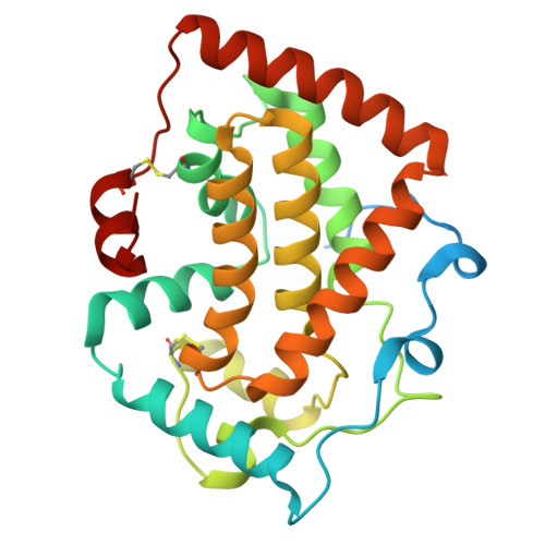
    9HTZ

  • PubMed Abstract: 

    M2-32 is a non-specific acid phosphatase with a rare ability to function across a broad pH range (3.5-8.5). Analysis using SWISS-PROT Prf Profiles classifies it as a class A acid phosphatase (Z-score: 78.97), sharing 50%-60% sequence similarity with enzymes such as PhoC and PhoN. For detailed characterization, the gene encoding M2-32 was cloned into the pET28(b) vector, overexpressed in Escherichia coli BL21 (DE3), and subsequently purified. Although the monomeric form of M2-32 has a molecular weight of ~28 kDa, size exclusion chromatography, dynamic light scattering, and sedimentation studies revealed a dimeric form in solution. Enzymatic assays using p-nitrophenyl phosphate, 4-methylumbelliferyl phosphate, 3'-and 5'-adenosine monophosphate demonstrated robust activity over a pH range of 4.0-8.0 at both 30 and 50°C. Differential scanning fluorimetry indicated an unfolding temperature close to 47°C; however, the enzyme refolded after heat denaturation at 80°C. We have determined the x-ray crystal structure of M2-32 by molecular replacement using an AlphaFold2-guided truncated model, achieving a resolution of 2.2 Å. The protein crystallized as a dimer-of-dimers. Each monomer (residues 38-274) adopts an all-alpha-helical fold composed of 14 helices and two disulfide bonds. Docking studies with adenosine monophosphates, combined with site-directed mutagenesis, identified His174, Arg207, His213, Asp217 as critical catalytic residues, and Tyr136 and Ser172 probably involved in substrate recognition. Mutations at these positions resulted in over 90% loss of enzymatic activity, highlighting their functional significance.


  • Organizational Affiliation
    • Estación Experimental del Zaidín, Consejo Superior de Investigaciones Científicas, Granada, Spain.

Macromolecules
Find similar proteins by:  (by identity cutoff)  |  3D Structure
Entity ID: 1
MoleculeChains Sequence LengthOrganismDetailsImage
Phosphatase PAP2 family protein
A, B, C, D
274metagenomeMutation(s): 0 
Gene Names: ABCR88_01690
EC: 3.1.3.2
UniProt
Find proteins for A0AAU7WWP3 (Pseudomonas sp. W17)
Explore A0AAU7WWP3 
Go to UniProtKB:  A0AAU7WWP3
Entity Groups  
Sequence Clusters30% Identity50% Identity70% Identity90% Identity95% Identity100% Identity
UniProt GroupA0AAU7WWP3
Sequence Annotations
Expand
  • Reference Sequence
Small Molecules
Ligands 5 Unique
IDChains Name / Formula / InChI Key2D Diagram3D Interactions
PEG
Query on PEG

Download Ideal Coordinates CCD File 
J [auth A],
K [auth A]
DI(HYDROXYETHYL)ETHER
C4 H10 O3
MTHSVFCYNBDYFN-UHFFFAOYSA-N
SO4
Query on SO4

Download Ideal Coordinates CCD File 
AA [auth C]
BA [auth C]
DA [auth D]
L [auth A]
M [auth A]
AA [auth C],
BA [auth C],
DA [auth D],
L [auth A],
M [auth A],
S [auth B],
T [auth B]
SULFATE ION
O4 S
QAOWNCQODCNURD-UHFFFAOYSA-L
GOL
Query on GOL

Download Ideal Coordinates CCD File 
H [auth A]
I [auth A]
P [auth B]
R [auth B]
V [auth C]
H [auth A],
I [auth A],
P [auth B],
R [auth B],
V [auth C],
Z [auth C]
GLYCEROL
C3 H8 O3
PEDCQBHIVMGVHV-UHFFFAOYSA-N
EDO
Query on EDO

Download Ideal Coordinates CCD File 
CA [auth D]
F [auth A]
G [auth A]
O [auth B]
U [auth C]
CA [auth D],
F [auth A],
G [auth A],
O [auth B],
U [auth C],
W [auth C],
X [auth C],
Y [auth C]
1,2-ETHANEDIOL
C2 H6 O2
LYCAIKOWRPUZTN-UHFFFAOYSA-N
ACT
Query on ACT

Download Ideal Coordinates CCD File 
E [auth A],
N [auth B],
Q [auth B]
ACETATE ION
C2 H3 O2
QTBSBXVTEAMEQO-UHFFFAOYSA-M
Experimental Data & Validation

Experimental Data

  • Method: X-RAY DIFFRACTION
  • Resolution: 2.20 Å
  • R-Value Free:  0.243 (Depositor), 0.244 (DCC) 
  • R-Value Work:  0.204 (Depositor), 0.205 (DCC) 
  • R-Value Observed: 0.205 (Depositor) 
Space Group: P 2 21 21
Unit Cell:
Length ( Å )Angle ( ˚ )
a = 61.07α = 90
b = 123.637β = 90
c = 138.646γ = 90
Software Package:
Software NamePurpose
PHENIXrefinement
Aimlessdata scaling
XDSdata reduction
MOLREPphasing

Structure Validation

View Full Validation Report


Entry History & Funding Information

Deposition Data


Funding OrganizationLocationGrant Number
Ministry of Economy and Competitiveness (MINECO)SpainPDI-2018-094370BI00
Ministry of Economy and Competitiveness (MINECO)SpainPID2020-116261GB-I00

Revision History  (Full details and data files)

  • Version 1.0: 2025-09-17
    Type: Initial release