9IN6 | pdb_00009in6

Capsid of Vibrio cholerae phage mature VP1

  • Classification: VIRAL PROTEIN
  • Organism(s): Vibrio cholerae
  • Mutation(s): No 

  • Deposited: 2024-07-05 Released: 2024-08-14 
  • Deposition Author(s): Liu, H.R., Pang, H.
  • Funding Organization(s): National Natural Science Foundation of China (NSFC)

Experimental Data Snapshot

  • Method: ELECTRON MICROSCOPY
  • Resolution: 2.80 Å
  • Aggregation State: PARTICLE 
  • Reconstruction Method: SINGLE PARTICLE 

wwPDB Validation   3D Report Full Report


This is version 1.3 of the entry. See complete history


Literature

Three-dimensional structures of Vibrio cholerae typing podophage VP1 in two states.

Pang, H.Fan, F.Zheng, J.Xiao, H.Tan, Z.Song, J.Kan, B.Liu, H.

(2024) Structure 32: 2364-2374.e2

  • DOI: https://doi.org/10.1016/j.str.2024.10.005
  • Primary Citation of Related Structures:  
    8ZKK, 8ZKM, 9IN6

  • PubMed Abstract: 

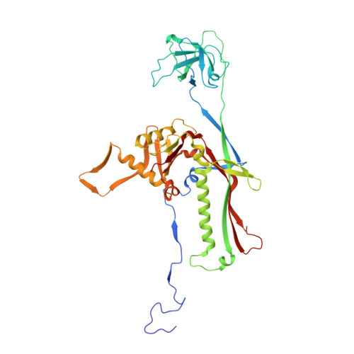
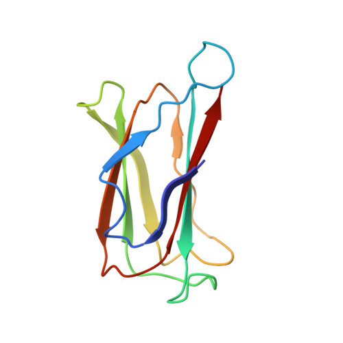
    Lytic podophages (VP1-VP5) play crucial roles in subtyping Vibrio cholerae O1 biotype El Tor. However, until now no structures of these phages have been available, which hindered our understanding of the molecular mechanisms of infection and DNA release. Here, we determined the cryoelectron microscopy (cryo-EM) structures of mature and DNA-ejected VP1 structures at near-atomic and subnanometer resolutions, respectively. The VP1 head is composed of 415 copies of the major capsid protein gp7 and 11 turret-shaped spikes. The VP1 tail consists of an adapter, a nozzle, a slender ring, and a tail needle, and is flanked by three extended fibers I and six trimeric fibers II. Conformational changes of fiber II in DNA-ejected VP1 may cause the release of the tail needle and core proteins, forming an elongated tail channel. Our structures provide insights into the molecular mechanisms of infection and DNA release for podophages with a tail needle.


  • Organizational Affiliation

    Institute of Interdisciplinary Studies, Key Laboratory for Matter Microstructure and Function of Hunan Province, Key Laboratory of Low-dimensional Quantum Structures and Quantum Control, School of Physics and Electronics, Hunan Normal University, Changsha 410082, China.


Macromolecules
Find similar proteins by:  (by identity cutoff)  |  3D Structure
Entity ID: 1
MoleculeChains Sequence LengthOrganismDetailsImage
major capsid of VP1399Vibrio choleraeMutation(s): 0 
Entity Groups  
Sequence Clusters30% Identity50% Identity70% Identity90% Identity95% Identity100% Identity
Sequence Annotations
Expand
  • Reference Sequence
Find similar proteins by:  (by identity cutoff)  |  3D Structure
Entity ID: 2
MoleculeChains Sequence LengthOrganismDetailsImage
SHP of VP1H [auth c]111Vibrio choleraeMutation(s): 0 
Entity Groups  
Sequence Clusters30% Identity50% Identity70% Identity90% Identity95% Identity100% Identity
Sequence Annotations
Expand
  • Reference Sequence
Experimental Data & Validation

Experimental Data

  • Method: ELECTRON MICROSCOPY
  • Resolution: 2.80 Å
  • Aggregation State: PARTICLE 
  • Reconstruction Method: SINGLE PARTICLE 
EM Software:
TaskSoftware PackageVersion
MODEL REFINEMENTPHENIX

Structure Validation

View Full Validation Report


Entry History & Funding Information

Deposition Data


Funding OrganizationLocationGrant Number
National Natural Science Foundation of China (NSFC)China12034006
National Natural Science Foundation of China (NSFC)China32071209
National Natural Science Foundation of China (NSFC)China32200994

Revision History  (Full details and data files)

  • Version 1.0: 2024-08-14
    Type: Initial release
  • Version 1.1: 2024-11-13
    Changes: Data collection, Database references, Structure summary
  • Version 1.2: 2024-12-18
    Changes: Data collection, Database references
  • Version 1.3: 2025-06-25
    Changes: Data collection