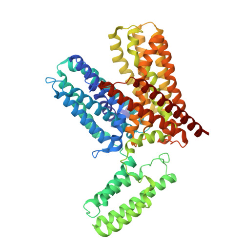
9J9Z | pdb_00009j9z

Cryo-EM structure of Outward state Anhydromuropeptide permease (AmpG) complex with GlcNAc-1,6-anhMurNAc


Experimental Data Snapshot

  • Method: ELECTRON MICROSCOPY
  • Resolution: 2.97 Å
  • Aggregation State: PARTICLE 
  • Reconstruction Method: SINGLE PARTICLE 

wwPDB Validation   3D Report Full Report


This is version 1.0 of the entry. See complete history


Literature

Structural and functional insights of AmpG in muropeptide transport and multiple beta-lactam antibiotics resistance.

Chang, N.Kim, H.Kim, U.Cho, Y.Yoo, Y.Lee, H.Kim, J.W.Kim, M.S.Lee, J.Cho, Y.L.Kim, K.Yong, D.Cho, H.S.

(2025) Nat Commun 16: 5744-5744

  • DOI: https://doi.org/10.1038/s41467-025-61169-3
  • Primary Citation of Related Structures:  
    8ZBB, 8ZGZ, 8ZKE, 9J9Z

  • PubMed Abstract: 

    Anhydromuropeptide permease (AmpG) is a transporter protein located in the inner membrane of certain gram -negative bacteria, involved in peptidoglycan (PG) recycling and β-lactamase induction. Decreased AmpG function reduces resistance of antibiotic-resistant bacteria to β-lactam antibiotics. Therefore, AmpG-targeting inhibitors are promising 'antibiotic adjuvants'. However, as the tertiary structure of AmpG has not yet been identified, the development of targeted inhibitors remains challenging. We present four cryo-electron microscopy (cryo-EM) structures: the apo-inward and apo-outward state structures and the inward-occluded and outward states complexed with the substrate GlcNAc-1,6-anhMurNAc. Through functional analysis and molecular dynamics (MD) simulations, we identified motif A, which stabilizes the outward state, substrate-binding pocket, and protonation-related residues. Based on the structure of AmpG and our experimental results, we propose a muropeptide transport mechanism for AmpG. A deeper understanding of its structure and transport mechanism provides a foundation for the development of antibiotic adjuvants.


  • Organizational Affiliation
    • Department of Systems Biology and Division of Life Sciences, Yonsei University, 50 Yonsei-ro, Seoul, Republic of Korea.

Macromolecules
Find similar proteins by:  (by identity cutoff)  |  3D Structure
Entity ID: 1
MoleculeChains Sequence LengthOrganismDetailsImage
anti-BRIL Fab Heavy chainA [auth H]225Homo sapiensMutation(s): 0 
Gene Names: IGHG1
Entity Groups  
Sequence Clusters30% Identity50% Identity70% Identity90% Identity95% Identity100% Identity
Sequence Annotations
Expand
  • Reference Sequence
Find similar proteins by:  (by identity cutoff)  |  3D Structure
Entity ID: 2
MoleculeChains Sequence LengthOrganismDetailsImage
anti-BRIL Fab NanobodyB [auth K]120synthetic constructMutation(s): 0 
Entity Groups  
Sequence Clusters30% Identity50% Identity70% Identity90% Identity95% Identity100% Identity
Sequence Annotations
Expand
  • Reference Sequence
Find similar proteins by:  (by identity cutoff)  |  3D Structure
Entity ID: 3
MoleculeChains Sequence LengthOrganismDetailsImage
anti-BRIL Fab Light chainC [auth L]212Homo sapiensMutation(s): 0 
Gene Names: IGHG1
Entity Groups  
Sequence Clusters30% Identity50% Identity70% Identity90% Identity95% Identity100% Identity
Sequence Annotations
Expand
  • Reference Sequence
Find similar proteins by:  (by identity cutoff)  |  3D Structure
Entity ID: 4
MoleculeChains Sequence LengthOrganismDetailsImage
Muropeptide transporter,Soluble cytochrome b562D [auth A]589Yokenella regensburgeiEscherichia coliMutation(s): 4 
Gene Names: ampGNCTC11967_01129cybC
Membrane Entity: Yes 
UniProt
Find proteins for A0AB38FS76 (Yokenella regensburgei)
Explore A0AB38FS76 
Go to UniProtKB:  A0AB38FS76
Entity Groups  
Sequence Clusters30% Identity50% Identity70% Identity90% Identity95% Identity100% Identity
UniProt GroupA0AB38FS76
Sequence Annotations
Expand
  • Reference Sequence
Small Molecules
Ligands 1 Unique
IDChains Name / Formula / InChI Key2D Diagram3D Interactions
2YP (Subject of Investigation/LOI)
Query on 2YP

Download Ideal Coordinates CCD File 
E [auth A](2R)-2-[[(1R,2S,3R,4R,5R)-4-acetamido-2-[(2S,3R,4R,5S,6R)-3-acetamido-6-(hydroxymethyl)-4,5-bis(oxidanyl)oxan-2-yl]oxy-6,8-dioxabicyclo[3.2.1]octan-3-yl]oxy]propanoic acid
C19 H30 N2 O12
MWWQKONGFKUAEK-STFZFCBQSA-N
Experimental Data & Validation

Experimental Data

  • Method: ELECTRON MICROSCOPY
  • Resolution: 2.97 Å
  • Aggregation State: PARTICLE 
  • Reconstruction Method: SINGLE PARTICLE 
EM Software:
TaskSoftware PackageVersion
MODEL REFINEMENTPHENIX1.21.2_5419

Structure Validation

View Full Validation Report


Entry History & Funding Information

Deposition Data


Funding OrganizationLocationGrant Number
Not funded--

Revision History  (Full details and data files)

  • Version 1.0: 2025-07-16
    Type: Initial release