9JR2 | pdb_00009jr2

Cryo-EM structure of PTH-PTH1R-Gq (upright state)


Experimental Data Snapshot

  • Method: ELECTRON MICROSCOPY
  • Resolution: 2.80 Å
  • Aggregation State: PARTICLE 
  • Reconstruction Method: SINGLE PARTICLE 

wwPDB Validation   3D Report Full Report


This is version 1.3 of the entry. See complete history


Literature

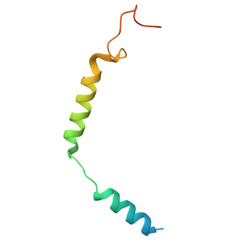
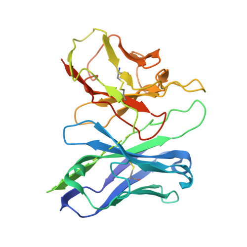
Insights into G-protein coupling preference from cryo-EM structures of G q -bound PTH1R.

Sano, F.K.Shimizume, K.Kobayashi, K.Awazu, T.Kawakami, K.Akasaka, H.Kobayashi, T.A.Tanaka, T.Okamoto, H.H.Hirano, H.Kusakizako, T.Shihoya, W.Kise, Y.Itoh, Y.Ishitani, R.Okada, Y.Sako, Y.Yanagawa, M.Inoue, A.Nureki, O.

(2025) Nat Chem Biol 

  • DOI: https://doi.org/10.1038/s41589-025-01957-6
  • Primary Citation of Related Structures:  
    9JR2, 9JR3

  • PubMed Abstract: 

    The parathyroid hormone type 1 receptor (PTH1R) is a prototypical class B1 G-protein-coupled receptor that couples to both G q and G s , having a crucial role in calcium homeostasis and serving as a therapeutic target for osteoporosis. Therapies targeting PTH1R face challenges because of G q -associated prolonged signaling, which leads to bone resorption. To address this, selective activation of G s signaling is desirable. However, the structural basis of G q -mediated signaling remains unclear, limiting the development of signal-selective drugs. Here, we present cryo-electron microscopy structures of the PTH1R-G q complex in two distinct extracellular conformations, demonstrating the role of N-linked glycans at N176 1.28 in stabilizing the ligand-tilted conformation. Comparison with a G s -bound PTH1R structure highlights the role of key interactions involving both the C terminus of Gα and the receptor's intracellular loop 2 in G q signaling. These structural insights provide a foundation for understanding the molecular mechanisms of PTH1R signaling.


  • Organizational Affiliation
    • Department of Biological Sciences, Graduate School of Science, The University of Tokyo, Tokyo, Japan.

Macromolecules
Find similar proteins by:  (by identity cutoff)  |  3D Structure
Entity ID: 1
MoleculeChains Sequence LengthOrganismDetailsImage
Guanine nucleotide-binding protein G(q) subunit alpha-1 (miniGq)245Homo sapiensMutation(s): 0 
EC: 3.6.5
Membrane Entity: Yes 
UniProt & NIH Common Fund Data Resources
Find proteins for P63092 (Homo sapiens)
Explore P63092 
Go to UniProtKB:  P63092
PHAROS:  P63092
GTEx:  ENSG00000087460 
Entity Groups  
Sequence Clusters30% Identity50% Identity70% Identity90% Identity95% Identity100% Identity
UniProt GroupP63092
Sequence Annotations
Expand
  • Reference Sequence
Find similar proteins by:  (by identity cutoff)  |  3D Structure
Entity ID: 2
MoleculeChains Sequence LengthOrganismDetailsImage
Guanine nucleotide-binding protein G(I)/G(S)/G(T) subunit beta-1344Rattus rattusMutation(s): 0 
Gene Names: Gnb1
UniProt
Find proteins for P54311 (Rattus norvegicus)
Explore P54311 
Go to UniProtKB:  P54311
Entity Groups  
Sequence Clusters30% Identity50% Identity70% Identity90% Identity95% Identity100% Identity
UniProt GroupP54311
Sequence Annotations
Expand
  • Reference Sequence
Find similar proteins by:  (by identity cutoff)  |  3D Structure
Entity ID: 3
MoleculeChains Sequence LengthOrganismDetailsImage
Guanine nucleotide-binding protein G(I)/G(S)/G(O) subunit gamma-2C [auth G]67Bos taurusMutation(s): 0 
Gene Names: GNG2
Membrane Entity: Yes 
UniProt
Find proteins for P63212 (Bos taurus)
Explore P63212 
Go to UniProtKB:  P63212
Entity Groups  
Sequence Clusters30% Identity50% Identity70% Identity90% Identity95% Identity100% Identity
UniProt GroupP63212
Sequence Annotations
Expand
  • Reference Sequence
Find similar proteins by:  (by identity cutoff)  |  3D Structure
Entity ID: 4
MoleculeChains Sequence LengthOrganismDetailsImage
scFv16D [auth S]260Mus musculusMutation(s): 0 
Entity Groups  
Sequence Clusters30% Identity50% Identity70% Identity90% Identity95% Identity100% Identity
Sequence Annotations
Expand
  • Reference Sequence
Find similar proteins by:  (by identity cutoff)  |  3D Structure
Entity ID: 5
MoleculeChains Sequence LengthOrganismDetailsImage
Parathyroid hormoneE [auth P]35Homo sapiensMutation(s): 1 
Membrane Entity: Yes 
UniProt & NIH Common Fund Data Resources
Find proteins for P01270 (Homo sapiens)
Explore P01270 
Go to UniProtKB:  P01270
PHAROS:  P01270
GTEx:  ENSG00000152266 
Entity Groups  
Sequence Clusters30% Identity50% Identity70% Identity90% Identity95% Identity100% Identity
UniProt GroupP01270
Sequence Annotations
Expand
  • Reference Sequence
Find similar proteins by:  (by identity cutoff)  |  3D Structure
Entity ID: 6
MoleculeChains Sequence LengthOrganismDetailsImage
Parathyroid hormone/parathyroid hormone-related peptide receptorF [auth R]567Homo sapiensMutation(s): 0 
Gene Names: PTH1RPTHRPTHR1
Membrane Entity: Yes 
UniProt & NIH Common Fund Data Resources
Find proteins for Q03431 (Homo sapiens)
Explore Q03431 
Go to UniProtKB:  Q03431
PHAROS:  Q03431
GTEx:  ENSG00000160801 
Entity Groups  
Sequence Clusters30% Identity50% Identity70% Identity90% Identity95% Identity100% Identity
UniProt GroupQ03431
Sequence Annotations
Expand
  • Reference Sequence
Experimental Data & Validation

Experimental Data

  • Method: ELECTRON MICROSCOPY
  • Resolution: 2.80 Å
  • Aggregation State: PARTICLE 
  • Reconstruction Method: SINGLE PARTICLE 
EM Software:
TaskSoftware PackageVersion
MODEL REFINEMENTPHENIX

Structure Validation

View Full Validation Report


Entry History & Funding Information

Deposition Data


Funding OrganizationLocationGrant Number
Japan Society for the Promotion of Science (JSPS)Japan--

Revision History  (Full details and data files)

  • Version 1.0: 2024-10-16
    Type: Initial release
  • Version 1.1: 2024-11-06
    Changes: Data collection
  • Version 1.2: 2025-07-02
    Changes: Data collection
  • Version 1.3: 2025-07-09
    Changes: Data collection, Database references