9L2N | pdb_00009l2n

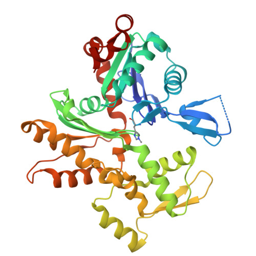
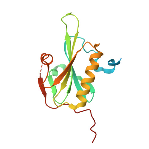
Crystal structure of Cytochalasin D bound to a filamentous conformation actin


Experimental Data Snapshot

  • Method: X-RAY DIFFRACTION
  • Resolution: 1.70 Å
  • R-Value Free: 
    0.204 (Depositor), 0.206 (DCC) 
  • R-Value Work: 
    0.175 (Depositor), 0.177 (DCC) 
  • R-Value Observed: 
    0.176 (Depositor) 

Starting Model: experimental
View more details

wwPDB Validation   3D Report Full Report


Ligand Structure Quality Assessment 


This is version 1.1 of the entry. See complete history


Literature

Microscopic and structural observations of actin filament capping and severing by cytochalasin D.

Mitani, T.Takeda, S.Oda, T.Narita, A.Maeda, Y.Honda, H.Fujiwara, I.

(2025) Proc Natl Acad Sci U S A 122: e2502164122-e2502164122

  • DOI: https://doi.org/10.1073/pnas.2502164122
  • Primary Citation of Related Structures:  
    9L2N

  • PubMed Abstract: 

    Cytochalasin D (CytoD), a widely used actin inhibitor, is typically employed in cell studies as a simple barbed end capper. However, accumulating evidence suggests broader effects on actin dynamics. We addressed this by observing single actin filaments using total internal reflection fluorescence microscopy. Our depolymerization assay confirmed that, at nanomolar concentrations, CytoD tightly caps barbed ends. The K 1/2 for inhibition was 4.1 nM, consistent with previous bulk measurements, and our approach revealed a capping duration of ~2 min. In polymerization assays, nanomolar CytoD completely suppressed barbed end elongation. Interestingly, at subnanomolar concentrations, CytoD caps barbed ends only transiently, rapidly associating and dissociating under both polymerizing and depolymerizing conditions. We interpreted this contradictory behavior as arising from differences in binding modes: capping one strand (fast dissociation) or both strands (slow dissociation). CytoD severs actin filaments at micromolar levels, a concentration range commonly used in cell biological studies. Although the severing rate is slower than cofilin, its higher frequency leads to filament fragmentation. Severing activity was suppressed by inorganic phosphate or cofilin. Our crystal structure of CytoD bound to filamentous conformation (F-form) actin showed that CytoD fits better in the hydrophobic cleft of F-form actin than monomeric conformation actin, explaining its preference for barbed end subunits. CytoD prevents barbed end depolymerization by stabilizing the terminal subunits in the F-form, which is supported by our MD simulations. These findings reveal the molecular mechanisms by which CytoD modulates actin dynamics and highlight the need for careful dosage control when treating cells with CytoD.


  • Organizational Affiliation

    Department of Materials Sciences and Bioengineering, Nagaoka University of Technology, Nagaoka, Niigata 940-2188, Japan.


Macromolecules
Find similar proteins by:  (by identity cutoff)  |  3D Structure
Entity ID: 1
MoleculeChains Sequence LengthOrganismDetailsImage
Actin, alpha skeletal muscle375Gallus gallusMutation(s): 0 
EC: 3.6.4
UniProt
Find proteins for P68139 (Gallus gallus)
Explore P68139 
Go to UniProtKB:  P68139
Entity Groups  
Sequence Clusters30% Identity50% Identity70% Identity90% Identity95% Identity100% Identity
UniProt GroupP68139
Sequence Annotations
Expand
  • Reference Sequence
Find similar proteins by:  (by identity cutoff)  |  3D Structure
Entity ID: 2
MoleculeChains Sequence LengthOrganismDetailsImage
Actin-binding protein fragmin P162Physarum polycephalumMutation(s): 1 
UniProt
Find proteins for Q94707 (Physarum polycephalum)
Explore Q94707 
Go to UniProtKB:  Q94707
Entity Groups  
Sequence Clusters30% Identity50% Identity70% Identity90% Identity95% Identity100% Identity
UniProt GroupQ94707
Sequence Annotations
Expand
  • Reference Sequence
Small Molecules
Ligands 6 Unique
IDChains Name / Formula / InChI Key2D Diagram3D Interactions
CY9 (Subject of Investigation/LOI)
Query on CY9

Download Ideal Coordinates CCD File 
H [auth A](3S,3aR,4S,6S,6aR,7E,10S,12R,13E,15R,15aR)-3-benzyl-6,12-dihydroxy-4,10,12-trimethyl-5-methylidene-1,11-dioxo-2,3,3a,4,5,6,6a,9,10,11,12,15-dodecahydro-1H-cycloundeca[d]isoindol-15-yl acetate
C30 H37 N O6
SDZRWUKZFQQKKV-JHADDHBZSA-N
ADP (Subject of Investigation/LOI)
Query on ADP

Download Ideal Coordinates CCD File 
C [auth A]ADENOSINE-5'-DIPHOSPHATE
C10 H15 N5 O10 P2
XTWYTFMLZFPYCI-KQYNXXCUSA-N
PO4
Query on PO4

Download Ideal Coordinates CCD File 
D [auth A],
F [auth A],
G [auth A]
PHOSPHATE ION
O4 P
NBIIXXVUZAFLBC-UHFFFAOYSA-K
EDO
Query on EDO

Download Ideal Coordinates CCD File 
K [auth B],
L [auth B]
1,2-ETHANEDIOL
C2 H6 O2
LYCAIKOWRPUZTN-UHFFFAOYSA-N
CA
Query on CA

Download Ideal Coordinates CCD File 
I [auth B],
J [auth B]
CALCIUM ION
Ca
BHPQYMZQTOCNFJ-UHFFFAOYSA-N
MG
Query on MG

Download Ideal Coordinates CCD File 
E [auth A]MAGNESIUM ION
Mg
JLVVSXFLKOJNIY-UHFFFAOYSA-N
Modified Residues  1 Unique
IDChains TypeFormula2D DiagramParent
HIC
Query on HIC
A
L-PEPTIDE LINKINGC7 H11 N3 O2HIS
Experimental Data & Validation

Experimental Data

  • Method: X-RAY DIFFRACTION
  • Resolution: 1.70 Å
  • R-Value Free:  0.204 (Depositor), 0.206 (DCC) 
  • R-Value Work:  0.175 (Depositor), 0.177 (DCC) 
  • R-Value Observed: 0.176 (Depositor) 
Space Group: P 21 21 21
Unit Cell:
Length ( Å )Angle ( ˚ )
a = 57.213α = 90
b = 90.806β = 90
c = 114.554γ = 90
Software Package:
Software NamePurpose
PHENIXrefinement
XDSdata scaling
XDSdata reduction
MOLREPphasing

Structure Validation

View Full Validation Report


Ligand Structure Quality Assessment 


Entry History & Funding Information

Deposition Data


Funding OrganizationLocationGrant Number
Japan Society for the Promotion of Science (JSPS)Japan16K17708
Japan Society for the Promotion of Science (JSPS)Japan17K07373
Japan Society for the Promotion of Science (JSPS)Japan20K06522
Japan Society for the Promotion of Science (JSPS)Japan22K06172
Japan Society for the Promotion of Science (JSPS)Japan23K05666

Revision History  (Full details and data files)

  • Version 1.0: 2025-07-02
    Type: Initial release
  • Version 1.1: 2025-07-30
    Changes: Database references