9LQL | pdb_00009lql

Structure of STG-hydrolyzing beta-glucosidase 1 (PSTG1) complexed with heptyl 1-thio-beta-D-glucopyranoside


Experimental Data Snapshot

  • Method: X-RAY DIFFRACTION
  • Resolution: 3.35 Å
  • R-Value Free: 
    0.286 (Depositor), 0.286 (DCC) 
  • R-Value Work: 
    0.245 (Depositor), 0.245 (DCC) 
  • R-Value Observed: 
    0.247 (Depositor) 

Starting Model: experimental
View more details

wwPDB Validation   3D Report Full Report


This is version 1.0 of the entry. See complete history


Literature

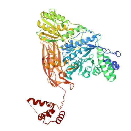
Structural and functional implications of the novel C-terminal domain and flexible loop in unique beta-glucosidase hydrolyzing sesaminol triglucoside to produce sesaminol

Yanai, T.Arai, A.Takahashi, Y.Imaizumi, R.Takeshita, K.Matsuura, H.Sakai, N.Takahashi, S.Yamamoto, M.Kataoka, K.Nakayama, T.Yamashita, S.

To be published.

Macromolecules
Find similar proteins by:  (by identity cutoff)  |  3D Structure
Entity ID: 1
MoleculeChains Sequence LengthOrganismDetailsImage
Beta-glucosidase
A, B, C, D
780Paenibacillus relictisesamiMutation(s): 0 
Gene Names: PSTG
EC: 3.2.1.21
UniProt
Find proteins for D6RVX0 (Paenibacillus relictisesami)
Explore D6RVX0 
Go to UniProtKB:  D6RVX0
Entity Groups  
Sequence Clusters30% Identity50% Identity70% Identity90% Identity95% Identity100% Identity
UniProt GroupD6RVX0
Sequence Annotations
Expand
  • Reference Sequence
Small Molecules
Ligands 4 Unique
IDChains Name / Formula / InChI Key2D Diagram3D Interactions
HTG (Subject of Investigation/LOI)
Query on HTG

Download Ideal Coordinates CCD File 
E [auth A],
H [auth B],
I [auth C]
heptyl 1-thio-beta-D-glucopyranoside
C13 H26 O5 S
HPEGNLMTTNTJSP-LBELIVKGSA-N
SO4
Query on SO4

Download Ideal Coordinates CCD File 
F [auth A],
J [auth C],
L [auth D]
SULFATE ION
O4 S
QAOWNCQODCNURD-UHFFFAOYSA-L
GOL (Subject of Investigation/LOI)
Query on GOL

Download Ideal Coordinates CCD File 
M [auth D]GLYCEROL
C3 H8 O3
PEDCQBHIVMGVHV-UHFFFAOYSA-N
EDO
Query on EDO

Download Ideal Coordinates CCD File 
G [auth B],
K [auth C]
1,2-ETHANEDIOL
C2 H6 O2
LYCAIKOWRPUZTN-UHFFFAOYSA-N
Experimental Data & Validation

Experimental Data

  • Method: X-RAY DIFFRACTION
  • Resolution: 3.35 Å
  • R-Value Free:  0.286 (Depositor), 0.286 (DCC) 
  • R-Value Work:  0.245 (Depositor), 0.245 (DCC) 
  • R-Value Observed: 0.247 (Depositor) 
Space Group: P 1
Unit Cell:
Length ( Å )Angle ( ˚ )
a = 64.6α = 71.503
b = 112.04β = 82.829
c = 156.59γ = 79.848
Software Package:
Software NamePurpose
PHENIXrefinement
XDSdata reduction
XDSdata scaling
PHASERphasing

Structure Validation

View Full Validation Report


Entry History & Funding Information

Deposition Data


Funding OrganizationLocationGrant Number
Japan Society for the Promotion of Science (JSPS)Japan24KJ1191
Japan Agency for Medical Research and Development (AMED)JapanJP20am0101070 (support number 2824)

Revision History  (Full details and data files)

  • Version 1.0: 2026-02-04
    Type: Initial release