


CD1c presenting phosphomycoketide in its open conformation
Cao, T.P., Rossjohn, J., Shahine, A.To be published.
Experimental Data Snapshot
Starting Model: experimental
View more details
Entity ID: 1 | |||||
|---|---|---|---|---|---|
| Molecule | Chains | Sequence Length | Organism | Details | Image |
| T-cell surface glycoprotein CD1c/T-cell surface glycoprotein CD1b chimeric protein | 278 | Homo sapiens | Mutation(s): 0 Gene Names: CD1C | 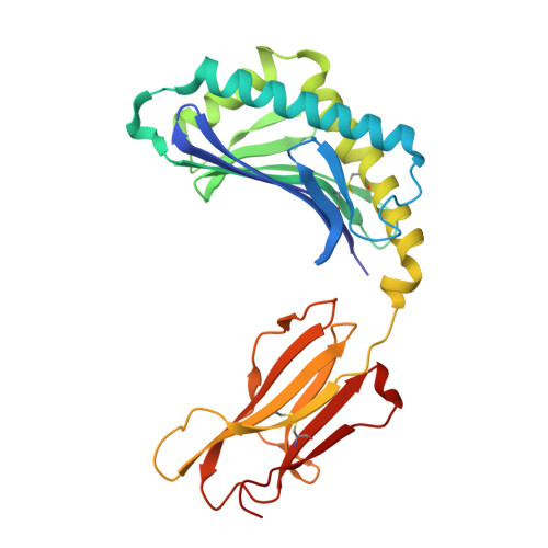 | |
Entity Groups | |||||
| Sequence Clusters | 30% Identity50% Identity70% Identity90% Identity95% Identity100% Identity | ||||
Glycosylation | |||||
| Glycosylation Sites: 1 | |||||
Sequence AnnotationsExpand | |||||
| |||||
Entity ID: 2 | |||||
|---|---|---|---|---|---|
| Molecule | Chains | Sequence Length | Organism | Details | Image |
| Beta-2-microglobulin | 98 | Homo sapiens | Mutation(s): 0 Gene Names: B2M, CDABP0092, HDCMA22P | 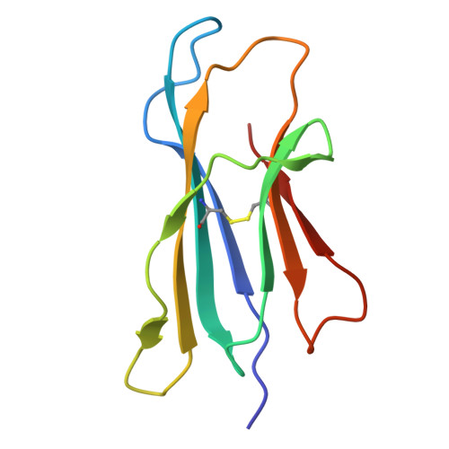 | |
UniProt & NIH Common Fund Data Resources | |||||
Find proteins for P61769 (Homo sapiens) Explore P61769 Go to UniProtKB: P61769 | |||||
PHAROS: P61769 GTEx: ENSG00000166710 | |||||
Entity Groups | |||||
| Sequence Clusters | 30% Identity50% Identity70% Identity90% Identity95% Identity100% Identity | ||||
| UniProt Group | P61769 | ||||
Sequence AnnotationsExpand | |||||
| |||||
| Ligands 6 Unique | |||||
|---|---|---|---|---|---|
| ID | Chains | Name / Formula / InChI Key | 2D Diagram | 3D Interactions | |
| A1CB8 (Subject of Investigation/LOI) Query on A1CB8 | D [auth A] | (4S,8S,12R,16S,20S)-4,8,12,16,20-pentamethylheptacosyl dihydrogen phosphate C32 H67 O4 P WLNOCCJMGSEDAT-LUKCZKMGSA-N |  | ||
| NAG Query on NAG | C [auth A] | 2-acetamido-2-deoxy-beta-D-glucopyranose C8 H15 N O6 OVRNDRQMDRJTHS-FMDGEEDCSA-N |  | ||
| KZF Query on KZF | G [auth A], H [auth A], I [auth A] | 2-(cyclohexylazaniumyl)ethanesulfonate C8 H17 N O3 S MKWKNSIESPFAQN-UHFFFAOYSA-N |  | ||
| GGG Query on GGG | J [auth A] | glycylglycylglycine C6 H11 N3 O4 XKUKSGPZAADMRA-UHFFFAOYSA-N |  | ||
| D12 Query on D12 | E [auth A], F [auth A] | DODECANE C12 H26 SNRUBQQJIBEYMU-UHFFFAOYSA-N |  | ||
| MLA Query on MLA | K [auth B], L [auth B] | MALONIC ACID C3 H4 O4 OFOBLEOULBTSOW-UHFFFAOYSA-N |  | ||
| Length ( Å ) | Angle ( ˚ ) |
|---|---|
| a = 54.452 | α = 90 |
| b = 83.468 | β = 90 |
| c = 92.541 | γ = 90 |
| Software Name | Purpose |
|---|---|
| PHENIX | refinement |
| PDB_EXTRACT | data extraction |
| ADSC | data collection |
| XDS | data reduction |
| Aimless | data scaling |
| PHASER | phasing |
| Funding Organization | Location | Grant Number |
|---|---|---|
| Australian Research Council (ARC) | Australia | DE210101031 |


