


Discovery of BBO-11818, a Potent and Selective Non-covalent Inhibitor of (ON) and (OFF) KRAS with Activity Against Multiple Oncogenic Mutants
Maciag, A.E., Chan, A.H., Simanshu, D.K., Beltran, P.J.(2025) Cancer Discov
Experimental Data Snapshot
Starting Model: experimental
View more details
(2025) Cancer Discov
Entity ID: 1 | |||||
|---|---|---|---|---|---|
| Molecule | Chains | Sequence Length | Organism | Details | Image |
| GTPase HRas | 170 | Homo sapiens | Mutation(s): 3 Gene Names: HRAS, HRAS1 EC: 3.6.5.2 | 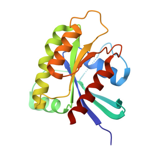 | |
UniProt & NIH Common Fund Data Resources | |||||
Find proteins for P01112 (Homo sapiens) Explore P01112 Go to UniProtKB: P01112 | |||||
PHAROS: P01112 GTEx: ENSG00000174775 | |||||
Entity Groups | |||||
| Sequence Clusters | 30% Identity50% Identity70% Identity90% Identity95% Identity100% Identity | ||||
| UniProt Group | P01112 | ||||
Sequence AnnotationsExpand | |||||
| |||||
| Ligands 3 Unique | |||||
|---|---|---|---|---|---|
| ID | Chains | Name / Formula / InChI Key | 2D Diagram | 3D Interactions | |
| A1CG4 (Subject of Investigation/LOI) Query on A1CG4 | E [auth A], H [auth B] | methyl (3S)-3-{[(7P)-7-(2-amino-3-cyano-7-fluoro-1-benzothiophen-4-yl)-8-fluoro-2-{[(2R,4R,7aS)-2-fluorotetrahydro-1H-pyrrolizin-7a(5H)-yl]methoxy}-6-(trifluoromethyl)quinazolin-4-yl](ethyl)amino}pyrrolidine-1-carboxylate C34 H33 F6 N7 O3 S VJMZMAQRBQSNHW-AXZPFCHASA-N |  | ||
| GNP Query on GNP | C [auth A], F [auth B] | PHOSPHOAMINOPHOSPHONIC ACID-GUANYLATE ESTER C10 H17 N6 O13 P3 UQABYHGXWYXDTK-UUOKFMHZSA-N |  | ||
| MG Query on MG | D [auth A], G [auth B] | MAGNESIUM ION Mg JLVVSXFLKOJNIY-UHFFFAOYSA-N |  | ||
| Length ( Å ) | Angle ( ˚ ) |
|---|---|
| a = 106.33 | α = 90 |
| b = 106.33 | β = 90 |
| c = 121.41 | γ = 120 |
| Software Name | Purpose |
|---|---|
| PHENIX | refinement |
| XSCALE | data scaling |
| XDS | data reduction |
| PHASER | phasing |
| PDB_EXTRACT | data extraction |
| Funding Organization | Location | Grant Number |
|---|---|---|
| National Institutes of Health/National Cancer Institute (NIH/NCI) | United States | 75N91019D00024 |


