9QEM | pdb_00009qem

Crystal structure of YTHDF2 in complex with compound 3 (AI-DF2-11)


Experimental Data Snapshot

  • Method: X-RAY DIFFRACTION
  • Resolution: 2.26 Å
  • R-Value Free: 
    0.260 (Depositor), 0.259 (DCC) 
  • R-Value Work: 
    0.219 (Depositor), 0.219 (DCC) 
  • R-Value Observed: 
    0.221 (Depositor) 

Starting Model: experimental
View more details

wwPDB Validation   3D Report Full Report


Ligand Structure Quality Assessment 


This is version 1.1 of the entry. See complete history


Literature

Discovery of YTHDF2 Ligands by Fragment-Based Design.

Invernizzi, A.Nai, F.Bedi, R.K.Vargas-Rosales, P.A.Li, Y.Bochenkova, E.Herok, M.Zalesak, F.Caflisch, A.

(2025) ACS Bio Med Chem Au 5: 753-765

  • DOI: https://doi.org/10.1021/acsbiomedchemau.5c00099
  • Primary Citation of Related Structures:  
    9QEL, 9QEM, 9QEO, 9QFL, 9QIU

  • PubMed Abstract: 

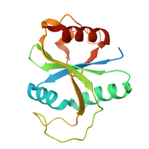
    N 6 -Adenosine methylation is the most abundant modification of mRNA. The three members of the YTH domain family proteins (YTHDF1-3) recognize in the cytoplasm the m 6 A-RNA modification. We screened a library of about 500,000 fragments (i.e., molecules with 11-20 non-hydrogen atoms) by docking into YTHDF2, which resulted in the identification of six active compounds among 47 tested in vitro (hit rate of 13%). The acquisition of 28 analogues of the docking hits provided an additional set of 10 active compounds (IC 50 < 100 μM). Protein crystallography-guided optimization of a ligand-efficient fragment by the synthesis of 32 derivatives culminated in a series of YTHDF2 ligands, which show low-micromolar affinity measured by a fluorescence polarization (FP) assay and a homogeneous time-resolved fluorescence-based (HTRF) assay. The series is characterized by very favorable ligand efficiency (of about 0.3-0.4 kcal/mol per non-hydrogen atom). Compound 23 binds to YTHDF2 according to the FP and HTRF assays with a K d value of 1.3 μM and an IC 50 value of 11 μM, respectively, and it is selective against all of the other YTH reader proteins. Several compounds of the series bind to the three YTHDF proteins with similar low-micromolar affinity, while they are less potent for YTHDC1 and YTHDC2. In contrast, compounds 17 and 30 bind also to YTHDC2, with K d of 6.3 and 4.9 μM, respectively. We also disclose six crystal structures of YTHDF2 in the complex with the fragments identified by docking.


  • Organizational Affiliation
    • Department of Biochemistry, University of Zurich, CH-8057 Zurich, Switzerland.

Macromolecules
Find similar proteins by:  (by identity cutoff)  |  3D Structure
Entity ID: 1
MoleculeChains Sequence LengthOrganismDetailsImage
YTH domain-containing family protein 2
A, B
167Homo sapiensMutation(s): 0 
Gene Names: YTHDF2HGRG8
UniProt & NIH Common Fund Data Resources
Find proteins for Q9Y5A9 (Homo sapiens)
Explore Q9Y5A9 
Go to UniProtKB:  Q9Y5A9
PHAROS:  Q9Y5A9
GTEx:  ENSG00000198492 
Entity Groups  
Sequence Clusters30% Identity50% Identity70% Identity90% Identity95% Identity100% Identity
UniProt GroupQ9Y5A9
Sequence Annotations
Expand
  • Reference Sequence
Small Molecules
Ligands 4 Unique
IDChains Name / Formula / InChI Key2D Diagram3D Interactions
A1I56 (Subject of Investigation/LOI)
Query on A1I56

Download Ideal Coordinates CCD File 
C [auth A],
I [auth B]
5-[[(3-methoxyphenyl)amino]methylidene]-2-sulfanylidene-1,3-diazinane-4,6-dione
C12 H11 N3 O3 S
TZPFBBAMHDIRBM-UHFFFAOYSA-N
SO4
Query on SO4

Download Ideal Coordinates CCD File 
F [auth A],
G [auth A],
J [auth B],
K [auth B],
L [auth B]
SULFATE ION
O4 S
QAOWNCQODCNURD-UHFFFAOYSA-L
GOL
Query on GOL

Download Ideal Coordinates CCD File 
D [auth A],
E [auth A]
GLYCEROL
C3 H8 O3
PEDCQBHIVMGVHV-UHFFFAOYSA-N
CL
Query on CL

Download Ideal Coordinates CCD File 
H [auth A]CHLORIDE ION
Cl
VEXZGXHMUGYJMC-UHFFFAOYSA-M
Experimental Data & Validation

Experimental Data

  • Method: X-RAY DIFFRACTION
  • Resolution: 2.26 Å
  • R-Value Free:  0.260 (Depositor), 0.259 (DCC) 
  • R-Value Work:  0.219 (Depositor), 0.219 (DCC) 
  • R-Value Observed: 0.221 (Depositor) 
Space Group: P 65
Unit Cell:
Length ( Å )Angle ( ˚ )
a = 80.06α = 90
b = 80.06β = 90
c = 110.8γ = 120
Software Package:
Software NamePurpose
PHENIXrefinement
XDSdata reduction
XSCALEdata scaling
PHASERphasing

Structure Validation

View Full Validation Report


Ligand Structure Quality Assessment 


Entry History & Funding Information

Deposition Data


Funding OrganizationLocationGrant Number
Swiss National Science FoundationSwitzerland310030-212195

Revision History  (Full details and data files)

  • Version 1.0: 2025-07-09
    Type: Initial release
  • Version 1.1: 2025-09-10
    Changes: Database references