


Crystal Structure of 6,7-dimethyl-8-ribityllumazine synthase from Bordetella pertussis in complex with 5-amino-6-(D-ribitylamino)uracil
Seibold, S., Lovell, S., Battaile, K.P.To be published.
Experimental Data Snapshot
Starting Model: in silico
View more details
Entity ID: 1 | |||||
|---|---|---|---|---|---|
| Molecule | Chains | Sequence Length | Organism | Details | Image |
| 6,7-dimethyl-8-ribityllumazine synthase | 163 | Bordetella pertussis Tohama I | Mutation(s): 0 Gene Names: ribH, BP3485 EC: 2.5.1.78 | 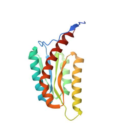 | |
UniProt | |||||
Find proteins for Q7VTN4 (Bordetella pertussis (strain Tohama I / ATCC BAA-589 / NCTC 13251)) Explore Q7VTN4 Go to UniProtKB: Q7VTN4 | |||||
Entity Groups | |||||
| Sequence Clusters | 30% Identity50% Identity70% Identity90% Identity95% Identity100% Identity | ||||
| UniProt Group | Q7VTN4 | ||||
Sequence AnnotationsExpand | |||||
| |||||
| Ligands 2 Unique | |||||
|---|---|---|---|---|---|
| ID | Chains | Name / Formula / InChI Key | 2D Diagram | 3D Interactions | |
| LMZ (Subject of Investigation/LOI) Query on LMZ | BA [auth E], L [auth A], P [auth B], T [auth C], X [auth D] | 5-NITROSO-6-RIBITYL-AMINO-2,4(1H,3H)-PYRIMIDINEDIONE C9 H14 N4 O7 YMWIHKCBRFEJMH-RPDRRWSUSA-N |  | ||
| CL Query on CL | AA [auth E] F [auth A] G [auth A] H [auth A] I [auth A] | CHLORIDE ION Cl VEXZGXHMUGYJMC-UHFFFAOYSA-M |  | ||
| Length ( Å ) | Angle ( ˚ ) |
|---|---|
| a = 97.126 | α = 90 |
| b = 75.061 | β = 105.57 |
| c = 180.117 | γ = 90 |
| Software Name | Purpose |
|---|---|
| PHENIX | refinement |
| Aimless | data scaling |
| XDS | data reduction |
| PHASER | phasing |
| PDB_EXTRACT | data extraction |
| Funding Organization | Location | Grant Number |
|---|---|---|
| National Institutes of Health/National Institute Of Allergy and Infectious Diseases (NIH/NIAID) | United States | 75N93022C00036 |
| National Institutes of Health/Office of the Director | United States | S10OD030394 |


