


Crystal structure of Glutamate--tRNA ligase (GltX) from Moraxella catarrhalis in complex with 5'-O-(N-Glutamyl)sulfamoyladeonosine
Enayati, P., Liu, L., Lovell, S., Battaile, K.P.To be published.
Experimental Data Snapshot
Starting Model: in silico
View more details
Entity ID: 1 | |||||
|---|---|---|---|---|---|
| Molecule | Chains | Sequence Length | Organism | Details | Image |
| Glutamate--tRNA ligase | 519 | Moraxella catarrhalis | Mutation(s): 0 Gene Names: gltX, AO370_0559 EC: 6.1.1.17 | 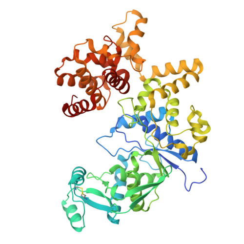 | |
UniProt | |||||
Find proteins for A0AB36DQE3 (Moraxella catarrhalis) Explore A0AB36DQE3 Go to UniProtKB: A0AB36DQE3 | |||||
Entity Groups | |||||
| Sequence Clusters | 30% Identity50% Identity70% Identity90% Identity95% Identity100% Identity | ||||
| UniProt Group | A0AB36DQE3 | ||||
Sequence AnnotationsExpand | |||||
| |||||
| Ligands 4 Unique | |||||
|---|---|---|---|---|---|
| ID | Chains | Name / Formula / InChI Key | 2D Diagram | 3D Interactions | |
| GSU (Subject of Investigation/LOI) Query on GSU | BA [auth C], E [auth A], IA [auth D], U [auth B] | O5'-(L-GLUTAMYL-SULFAMOYL)-ADENOSINE C15 H21 N7 O9 S YBRKRYFZKHICLS-WERHYGNASA-N |  | ||
| 2PE Query on 2PE | JA [auth D] | NONAETHYLENE GLYCOL C18 H38 O10 YZUUTMGDONTGTN-UHFFFAOYSA-N |  | ||
| P6G Query on P6G | V [auth B] | HEXAETHYLENE GLYCOL C12 H26 O7 IIRDTKBZINWQAW-UHFFFAOYSA-N |  | ||
| SO4 Query on SO4 | AA [auth B] CA [auth C] DA [auth C] EA [auth C] F [auth A] | SULFATE ION O4 S QAOWNCQODCNURD-UHFFFAOYSA-L |  | ||
| Length ( Å ) | Angle ( ˚ ) |
|---|---|
| a = 53.713 | α = 90 |
| b = 124.225 | β = 90 |
| c = 458.212 | γ = 90 |
| Software Name | Purpose |
|---|---|
| PHENIX | refinement |
| Aimless | data scaling |
| XDS | data reduction |
| PHASER | phasing |
| PDB_EXTRACT | data extraction |
| Funding Organization | Location | Grant Number |
|---|---|---|
| National Institutes of Health/National Institute Of Allergy and Infectious Diseases (NIH/NIAID) | United States | 75N93022C00036 |
| National Institutes of Health/Office of the Director | United States | S10OD030394 |


