10ZI | pdb_000010zi

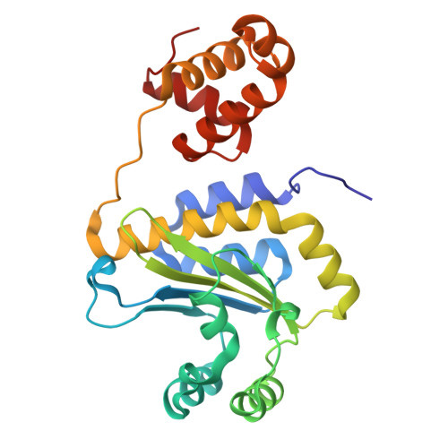
Crystal structure of MrtR bound to 3O-C8 homoserine lactone and tetraethylene glycol


Experimental Data Snapshot

  • Method: X-RAY DIFFRACTION
  • Resolution: 1.22 Å
  • R-Value Free: 
    0.193 (Depositor), 0.193 (DCC) 
  • R-Value Work: 
    0.155 (Depositor), 0.154 (DCC) 
  • R-Value Observed: 
    0.157 (Depositor) 

Starting Model: in silico
View more details

wwPDB Validation 3D Report Full Report

Validation slider image for 10ZI

Ligand Structure Quality Assessment 


This is version 1.1 of the entry. See complete history. 

Literature

MrtR of Mesorhizobium tianshanense reveals both activation and inhibition mechanisms of a LuxR-type quorum sensing receptor.

Stoutland, I.M., Blackwell, H.E.

(2026) Proc Natl Acad Sci U S A 123: e2616692123-e2616692123

  • DOI: https://doi.org/10.1073/pnas.2616692123
  • Primary Citation Related Structures: 
    10ZI, 11BM, 9Y2H, 9ZPJ, 9ZZ0

  • PubMed Abstract: 

    Quorum sensing (QS) enables common gram-negative bacteria to coordinate collective behaviors through small molecule signals, yet how these signals tune receptor activity remains incompletely understood. Here, we define a mechanism by which ligand structure controls function in a LuxR-type QS receptor. Using structural and biochemical analyses, we investigate MrtR from Mesorhizobium tianshanense and show that ligand acyl-chain length governs receptor assembly and activity. We present full-length structures of MrtR bound to activating and inhibitory ligands, revealing a switch in oligomeric state. Long-chain (C14) N -acyl l-homoserine lactones (AHLs) act as agonists by promoting intra- and intersubunit interactions that lead to homodimerization and DNA binding. In contrast, shorter (C8) AHLs fail to promote these contacts, favoring a monomeric, inactive state. Ligands of intermediate length produce graded responses consistent with partial dimer stabilization. Biochemical measurements of DNA binding, thermostability, and oligomerization, together with targeted mutagenesis, support this model and establish the functional importance of key structural contacts. These findings provide a side-by-side structural comparison of a full-length LuxR-type receptor bound to both agonist and antagonist. Our results expand the known structural and mechanistic diversity of the LuxR family and suggest mechanistic similarities between structurally distinct receptors.


  • Organizational Affiliation: 
    • Department of Chemistry, University of Wisconsin-Madison, Madison, WI 53706.

Macromolecule Content 

  • Total Structure Weight: 28.14 kDa 
  • Atom Count: 2,751 
  • Modeled Residue Count: 241 
  • Deposited Residue Count: 241 
  • Unique protein chains: 1

Macromolecules

Find similar proteins by:|  3D Structure
Entity ID: 1
MoleculeChains  Sequence LengthOrganismDetailsImage
MrtR241Mesorhizobium tianshanenseMutation(s): 0 
Gene Names: mrtR
UniProt
Find proteins for Q45NF4 (Mesorhizobium tianshanense)
Explore Q45NF4 
Go to UniProtKB:  Q45NF4
Entity Groups
Sequence Clusters30% Identity50% Identity70% Identity90% Identity95% Identity100% Identity
UniProt GroupQ45NF4
Sequence Annotations
Expand
Reference Sequence

Small Molecules

Ligands 5 Unique
IDChains Name / Formula / InChI Key2D Diagram3D Interactions
LAE
(Subject of Investigation/LOI)

Query on LAE


Download:Ideal Coordinates CCD File
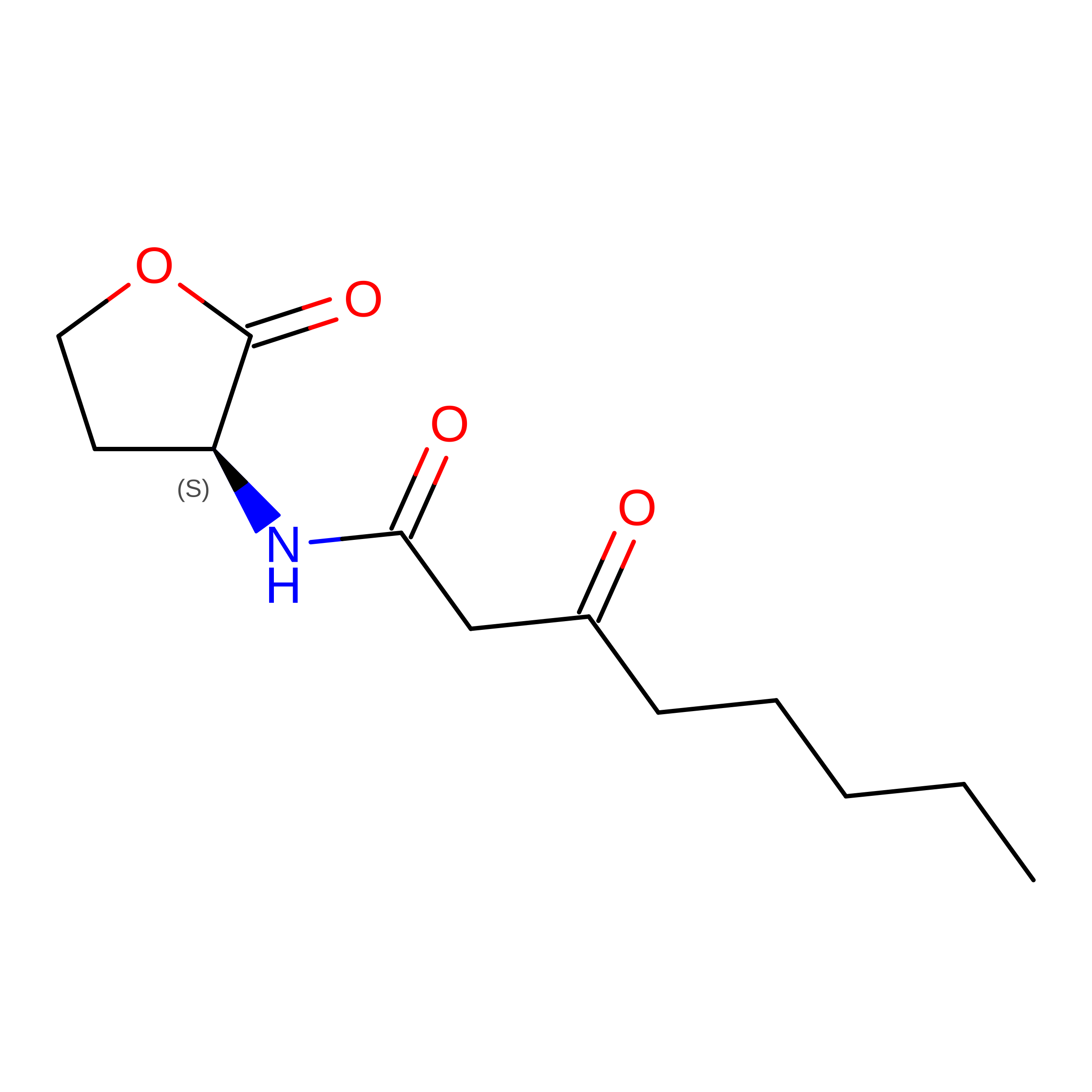
E [auth A]3-OXO-OCTANOIC ACID (2-OXO-TETRAHYDRO-FURAN-3-YL)-AMIDE
C12 H19 N O4
FXCMGCFNLNFLSH-JTQLQIEISA-N
PG4

Query on PG4


Download:Ideal Coordinates CCD File
G [auth A]TETRAETHYLENE GLYCOL
C8 H18 O5
UWHCKJMYHZGTIT-UHFFFAOYSA-N
MRD

Query on MRD


Download:Ideal Coordinates CCD File
C [auth A],
D [auth A]
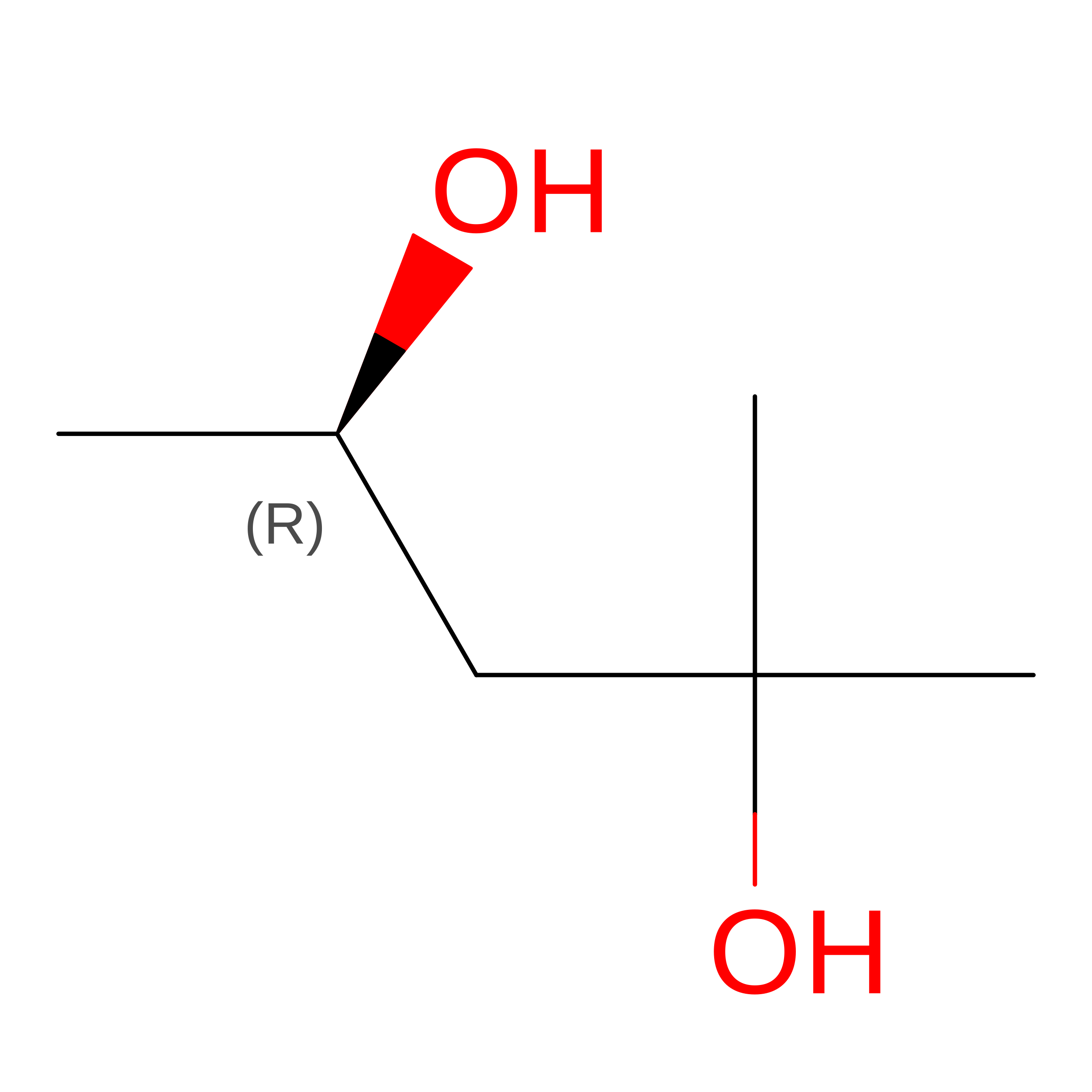
(4R)-2-METHYLPENTANE-2,4-DIOL
C6 H14 O2
SVTBMSDMJJWYQN-RXMQYKEDSA-N
MPD

Query on MPD


Download:Ideal Coordinates CCD File
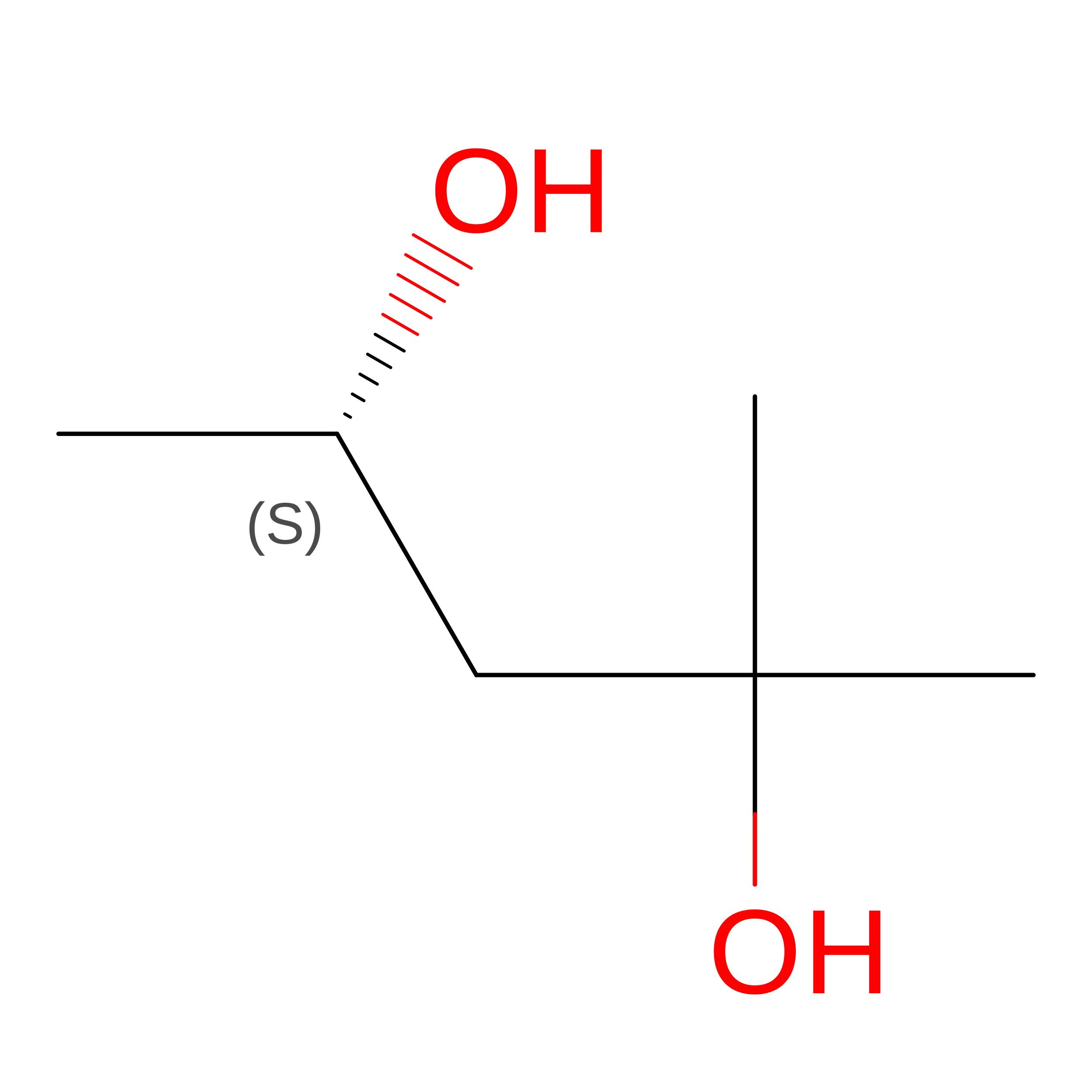
B [auth A](4S)-2-METHYL-2,4-PENTANEDIOL
C6 H14 O2
SVTBMSDMJJWYQN-YFKPBYRVSA-N
DMS

Query on DMS


Download:Ideal Coordinates CCD File
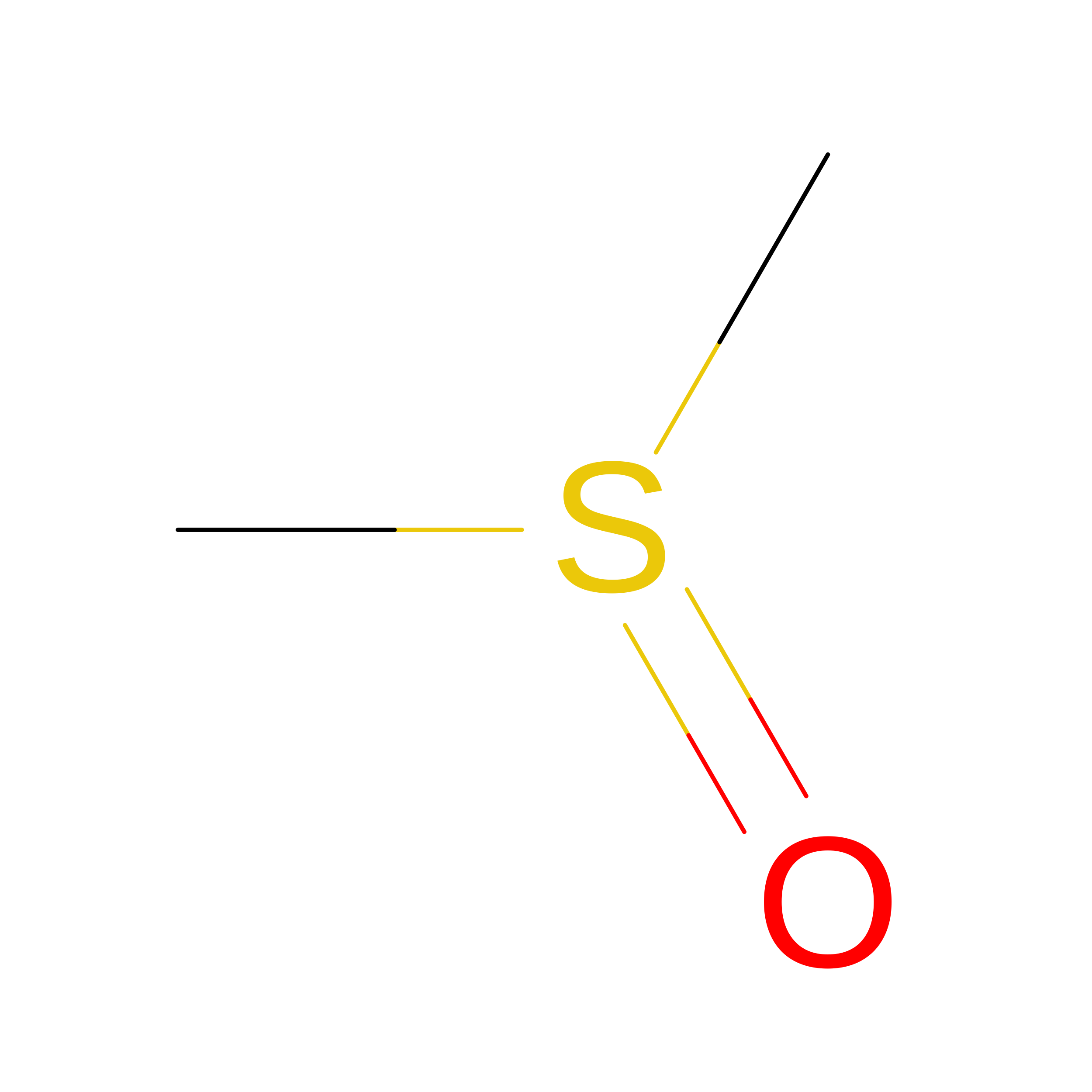
F [auth A]DIMETHYL SULFOXIDE
C2 H6 O S
IAZDPXIOMUYVGZ-UHFFFAOYSA-N

Experimental Data & Validation

Experimental Data

  • Method: X-RAY DIFFRACTION
  • Resolution: 1.22 Å
  • R-Value Free:  0.193 (Depositor), 0.193 (DCC) 
  • R-Value Work:  0.155 (Depositor), 0.154 (DCC) 
  • R-Value Observed: 0.157 (Depositor) 
Space Group: C 1 2 1
Unit Cell:
Length ( Å )Angle ( ˚ )
a = 126.434α = 90
b = 35.728β = 112.13
c = 61.941γ = 90
Software Package:
Software NamePurpose
REFMACrefinement
autoPROCdata reduction
autoPROCdata scaling
PHASERphasing

Structure Validation

View Full Validation Report


Ligand Structure Quality Assessment 


Entry History 

& Funding Information

Deposition Data


Funding OrganizationLocationGrant Number
National Institutes of Health/National Institute of General Medical Sciences (NIH/NIGMS)United States2R35GM131817-06

Revision History  (Full details and data files)

  • Version 1.0: 2026-09-23
    Type: Initial release
  • Version 1.1: 2026-10-14
    Changes: Database references