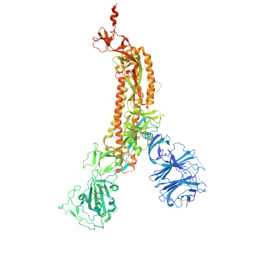
11UK | pdb_000011uk

Structure of SARS-CoV-2 variant LP.8.1 spike protein


Experimental Data Snapshot

  • Method: ELECTRON MICROSCOPY
  • Resolution: 2.64 Å
  • Aggregation State: PARTICLE 
  • Reconstruction Method: SINGLE PARTICLE 

wwPDB Validation 3D Report Full Report

Validation slider image for 11UK

This is version 1.0 of the entry. See complete history. 

Literature

mRNA-1273.251 and mRNA-1283.251 vaccines expressing SARS-CoV-2 variant LP.8.1 antigens broadly neutralize JN.1-lineage viruses

Nasir, A., Lee, D.W., Wang, Z., Ogega, C., Bopp, N., Wisti, P., Makrinos, D., Jani, H., Gorrie, A., Bender, M.F., Laciak, A.R., Sharma, A., Hu, X., Speidel, T., Ghantous, F., Berrueta, D.M., Hou, Y.J., Severson, M., Langley, A., Hardcastle, K., Elbashir, S., Harmon, C., Amato, N.J., Wu, K., Schief, W.R., Hohn, K., Carfi, A., Budigi, Y., Wahid, R., Edwards, D.K., Freyn, A.W.

(2026) Mol Ther Nucleic Acids 

Macromolecule Content 

  • Total Structure Weight: 435.26 kDa 
  • Atom Count: 25,353 
  • Modeled Residue Count: 3,099 
  • Deposited Residue Count: 3,777 
  • Unique protein chains: 1

Macromolecules

Find similar proteins by:|  3D Structure
Entity ID: 1
MoleculeChains  Sequence LengthOrganismDetailsImage
Spike glycoprotein
A, B, C
1,259Severe acute respiratory syndrome coronavirus 2Mutation(s): 0 
Entity Groups
Sequence Clusters30% Identity50% Identity70% Identity90% Identity95% Identity100% Identity
Glycosylation
Glycosylation Sites: 18
Sequence Annotations
Expand
Reference Sequence

Oligosaccharides

Help  
Entity ID: 2
MoleculeChains Length2D Diagram GlycosylationD Interactions
beta-D-mannopyranose-(1-4)-2-acetamido-2-deoxy-beta-D-glucopyranose-(1-4)-2-acetamido-2-deoxy-beta-D-glucopyranose
D, E, I, J, N
D, E, I, J, N, O
3N-Glycosylation
Entity ID: 3
MoleculeChains Length2D Diagram GlycosylationD Interactions
2-acetamido-2-deoxy-beta-D-glucopyranose-(1-4)-2-acetamido-2-deoxy-beta-D-glucopyranose
F, G, H, K, L
F, G, H, K, L, M, P, Q, R
2N-Glycosylation

Small Molecules

Ligands 1 Unique
IDChains Name / Formula / InChI Key2D Diagram3D Interactions
NAG

Query on NAG


Download:Ideal Coordinates CCD File
AA [auth A]
AB [auth C]
BA [auth A]
BB [auth C]
CA [auth A]
AA [auth A],
AB [auth C],
BA [auth A],
BB [auth C],
CA [auth A],
CB [auth C],
DA [auth A],
DB [auth C],
EA [auth A],
EB [auth C],
FA [auth B],
GA [auth B],
HA [auth B],
IA [auth B],
JA [auth B],
KA [auth B],
LA [auth B],
MA [auth B],
NA [auth B],
OA [auth B],
PA [auth B],
QA [auth B],
RA [auth B],
S [auth A],
SA [auth C],
T [auth A],
TA [auth C],
U [auth A],
UA [auth C],
V [auth A],
VA [auth C],
W [auth A],
WA [auth C],
X [auth A],
XA [auth C],
Y [auth A],
YA [auth C],
Z [auth A],
ZA [auth C]
2-acetamido-2-deoxy-beta-D-glucopyranose
C8 H15 N O6
OVRNDRQMDRJTHS-FMDGEEDCSA-N

Experimental Data & Validation

Experimental Data

  • Method: ELECTRON MICROSCOPY
  • Resolution: 2.64 Å
  • Aggregation State: PARTICLE 
  • Reconstruction Method: SINGLE PARTICLE 
EM Software:
TaskSoftware PackageVersion
RECONSTRUCTIONcryoSPARCv4.7.1

Structure Validation

View Full Validation Report


Entry History 

& Funding Information

Deposition Data


Funding OrganizationLocationGrant Number
Not funded--

Revision History  (Full details and data files)

  • Version 1.0: 2026-11-18
    Type: Initial release