Download Mendeley
Noncovalent SARS-CoV-2 main protease inhibitors targeting the catalytic dyad and primed substrate binding subsites.
Bhandari, D., Kovalevskaya, K., Coates, L., Jonsson, C.B., Parvathareddy, J., Weiss, K.L., Aniana, A., Louis, J.M., Bonnesen, P.V., Kovalevsky, A.(2026) RSC Med Chem
- PubMed: 42657162 Search on PubMedSearch on PubMed Central
- DOI: https://doi.org/10.1039/d6md00401f
- Primary Citation Related Structures:
12QH, 12SV, 12TA, 12WC, 12XD, 12YF, 12ZG, 12ZL, 13BW, 13CL, 13FA, 13HO - PubMed Abstract:
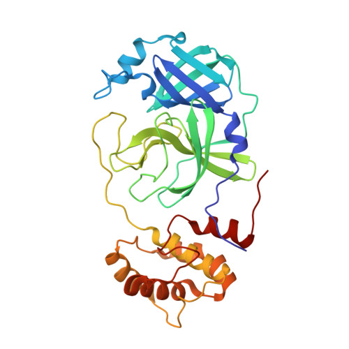
SARS-CoV-2 main protease (MPro) is a proven target for drug discovery of small-molecule antiviral agents due to its crucial role in viral polyprotein processing, high structural conservation across numerous divergent variants, and the lack of similar human enzymes. Unlike covalent compounds, noncovalent inhibitors of MPro do not modify the enzyme's active site, and may offer improved safety profiles, greater chemical tractability, and better oral bioavailability without the need for pharmacokinetic enhancement. In this study, we designed, synthesized and characterized thirteen noncovalent nonpeptidic SARS-CoV-2 MPro inhibitors clustered into two series (KK and KB) of compounds. The inhibitors were designed based on our recently discovered Mcule-5948770040 and its analogue HL-3-68 designed through the structure-activity relationship study. To obtain atomic details of the inhibitors' binding we solved room-temperature X-ray structures of the MPro/inhibitor complexes, and to quantify their binding and antiviral properties we performed in vitro DSF and ITC measurements and TCID 50 antiviral assays. In addition, a room-temperature neutron structure of the MPro/KB-5 complex allowed direct determination of hydrogen positions, mapping intermolecular interactions and directly visualizing the protonation states and hydrogen bonding. Improved binding affinities of KK-7 and KB-3 through KB-6 could be attributed to the observed nonconventional S-H⋯F hydrogen bond and an additional conventional hydrogen bond between the carboxamide moieties and Q189. Our study provides binding details for the designed compounds and demonstrates the feasibility of our joint X-ray/neutron structure-assisted drug design approach to generate more potent noncovalent nonpeptidic MPro inhibitors.
- Neutron Scattering Division, Oak Ridge National Laboratory Oak Ridge TN 37831 USA kovalevskyay@ornl.gov.
Organizational Affiliation: