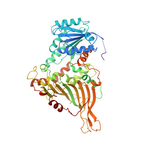
1QKI | pdb_00001qki

X-RAY STRUCTURE OF HUMAN GLUCOSE 6-PHOSPHATE DEHYDROGENASE (VARIANT CANTON R459L) COMPLEXED WITH STRUCTURAL NADP+


Experimental Data Snapshot

  • Method: X-RAY DIFFRACTION
  • Resolution: 3.00 Å
  • R-Value Free: 
    0.294 (Depositor) 
  • R-Value Work: 
    0.247 (Depositor), 0.240 (DCC) 
  • R-Value Observed: 
    0.247 (Depositor) 

Starting Model: experimental
View more details

wwPDB Validation   3D Report Full Report


Ligand Structure Quality Assessment 


This is version 2.1 of the entry. See complete history


Literature

Human Glucose-6-Phosphate Dehydrogenase: The Crystal Structure Reveals a Structural Nadp+ Molecule and Provides Insights Into Enzyme Deficiency

Au, S.W.N.Gover, S.Lam, V.M.S.Adams, M.J.

(2000) Structure 8: 293

  • DOI: https://doi.org/10.1016/s0969-2126(00)00104-0
  • Primary Citation of Related Structures:  
    1QKI

  • PubMed Abstract: 

    Glucose-6-phosphate dehydrogenase (G6PD) catalyses the first committed step in the pentose phosphate pathway; the generation of NADPH by this enzyme is essential for protection against oxidative stress. The human enzyme is in a dimer<-->tetramer equilibrium and its stability is dependent on NADP(+) concentration. G6PD deficiency results from many different point mutations in the X-linked gene encoding G6PD and is the most common human enzymopathy. Severe deficiency causes chronic non-spherocytic haemolytic anaemia; the usual symptoms are neonatal jaundice, favism and haemolytic anaemia. We have determined the first crystal structure of a human G6PD (the mutant Canton, Arg459-->Leu) at 3 A resolution. The tetramer is a dimer of dimers. Despite very similar dimer topology, there are two major differences from G6PD of Leuconostoc mesenteroides: a structural NADP(+) molecule, close to the dimer interface but integral to the subunit, is visible in all subunits of the human enzyme; and an intrasubunit disulphide bond tethers the otherwise disordered N-terminal segment. The few dimer-dimer contacts making the tetramer are charge-charge interactions. The importance of NADP(+) for stability is explained by the structural NADP(+) site, which is not conserved in prokaryotes. The structure shows that point mutations causing severe deficiency predominate close to the structural NADP(+) and the dimer interface, primarily affecting the stability of the molecule. They also indicate that a stable dimer is essential to retain activity in vivo. As there is an absolute requirement for some G6PD activity, residues essential for coenzyme or substrate binding are rarely modified.


  • Organizational Affiliation
    • Laboratory of Molecular Biophysics, Department of Biochemistry, University of Oxford, The University of Hong Kong, Department of Biochemistry, Oxford, OX1 3QU, UK, Hong Kong.

Macromolecules
Find similar proteins by:  (by identity cutoff)  |  3D Structure
Entity ID: 1
MoleculeChains Sequence LengthOrganismDetailsImage
GLUCOSE-6-PHOSPHATE 1-DEHYDROGENASE
A, B, C, D, E
A, B, C, D, E, F, G, H
514Homo sapiensMutation(s): 1 
Gene Names: G6PD
EC: 1.1.1.49
UniProt & NIH Common Fund Data Resources
Find proteins for P11413 (Homo sapiens)
Explore P11413 
Go to UniProtKB:  P11413
PHAROS:  P11413
GTEx:  ENSG00000160211 
Entity Groups  
Sequence Clusters30% Identity50% Identity70% Identity90% Identity95% Identity100% Identity
UniProt GroupP11413
Sequence Annotations
Expand
  • Reference Sequence
Small Molecules
Ligands 3 Unique
IDChains Name / Formula / InChI Key2D Diagram3D Interactions
NAP
Query on NAP

Download Ideal Coordinates CCD File 
BA [auth H]
I [auth A]
K [auth B]
O [auth C]
R [auth D]
BA [auth H],
I [auth A],
K [auth B],
O [auth C],
R [auth D],
T [auth E],
W [auth F],
Y [auth G]
NADP NICOTINAMIDE-ADENINE-DINUCLEOTIDE PHOSPHATE
C21 H28 N7 O17 P3
XJLXINKUBYWONI-NNYOXOHSSA-N
GOL
Query on GOL

Download Ideal Coordinates CCD File 
AA [auth G],
M [auth B],
N [auth B],
Q [auth C],
V [auth E]
GLYCEROL
C3 H8 O3
PEDCQBHIVMGVHV-UHFFFAOYSA-N
GOA
Query on GOA

Download Ideal Coordinates CCD File 
CA [auth H]
J [auth A]
L [auth B]
P [auth C]
S [auth D]
CA [auth H],
J [auth A],
L [auth B],
P [auth C],
S [auth D],
U [auth E],
X [auth F],
Z [auth G]
GLYCOLIC ACID
C2 H4 O3
AEMRFAOFKBGASW-UHFFFAOYSA-N
Experimental Data & Validation

Experimental Data

  • Method: X-RAY DIFFRACTION
  • Resolution: 3.00 Å
  • R-Value Free:  0.294 (Depositor) 
  • R-Value Work:  0.247 (Depositor), 0.240 (DCC) 
  • R-Value Observed: 0.247 (Depositor) 
Space Group: P 21 21 21
Unit Cell:
Length ( Å )Angle ( ˚ )
a = 128.9α = 90
b = 208.7β = 90
c = 214.3γ = 90
Software Package:
Software NamePurpose
X-PLORrefinement
DENZOdata reduction
SCALAdata scaling
X-PLORphasing

Structure Validation

View Full Validation Report


Ligand Structure Quality Assessment 


Entry History 

Deposition Data

Revision History  (Full details and data files)

  • Version 1.0: 2000-03-16
    Type: Initial release
  • Version 1.1: 2011-05-08
    Changes: Version format compliance
  • Version 1.2: 2011-07-13
    Changes: Version format compliance
  • Version 1.3: 2019-05-08
    Changes: Data collection, Experimental preparation, Other
  • Version 1.4: 2019-05-22
    Changes: Data collection, Refinement description
  • Version 2.0: 2023-11-15
    Changes: Atomic model, Data collection, Database references, Derived calculations, Other
  • Version 2.1: 2023-12-13
    Changes: Refinement description