
 Download Mendeley
Crystal structure of a variant c-di-GMP-II Riboswitch BhP1-5delC A9G/U55C bound with 2',3'-cGAMP
Xu, X.C., Ren, A.M.To be published.
Experimental Data Snapshot
Starting Model: experimental
View more details
Entity ID: 1 | ||||
| Molecule | Chains | Length | Organism | Image |
|---|---|---|---|---|
| RNA (77-MER) | 77 | Halalkalibacterium halodurans C-125 | 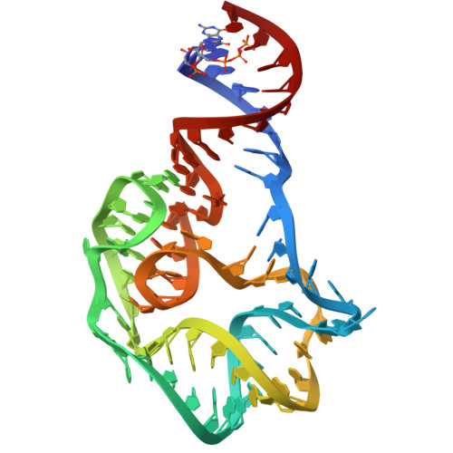 | |
Sequence AnnotationsExpand | ||||
Reference Sequence | ||||
| Ligands 2 Unique | |||||
|---|---|---|---|---|---|
| ID | Chains | Name / Formula / InChI Key | 2D Diagram | 3D Interactions | |
| 1SY (Subject of Investigation/LOI) Download:Ideal Coordinates CCD File | C [auth A], F [auth B] | cGAMP C20 H24 N10 O13 P2 XRILCFTWUCUKJR-INFSMZHSSA-N |  | ||
| MG Download:Ideal Coordinates CCD File | D [auth A], E [auth A], G [auth B] | MAGNESIUM ION Mg JLVVSXFLKOJNIY-UHFFFAOYSA-N |  | ||
| Length ( Å ) | Angle ( ˚ ) |
|---|---|
| a = 93.228 | α = 90 |
| b = 93.228 | β = 90 |
| c = 102.372 | γ = 120 |
| Software Name | Purpose |
|---|---|
| PHENIX | refinement |
| HKL-3000 | data scaling |
| PDB_EXTRACT | data extraction |
| HKL-3000 | data reduction |
| PHASER | phasing |
| Funding Organization | Location | Grant Number |
|---|---|---|
| National Natural Science Foundation of China (NSFC) | China | -- |
