22VG | pdb_000022vg

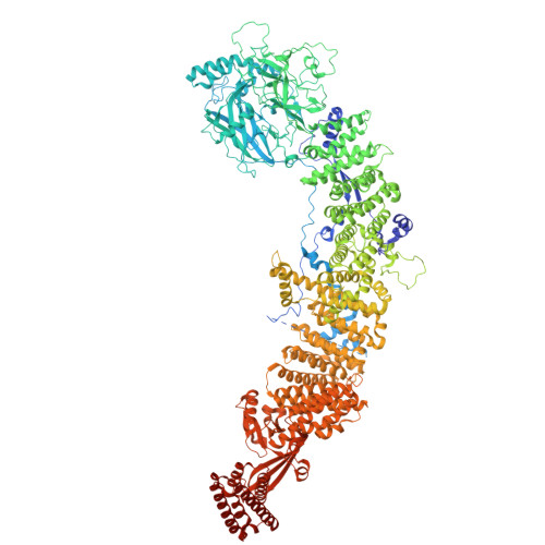
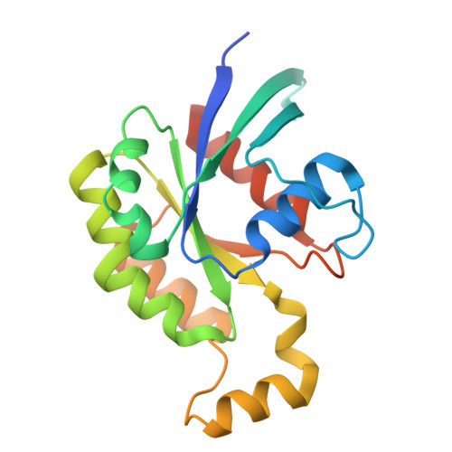
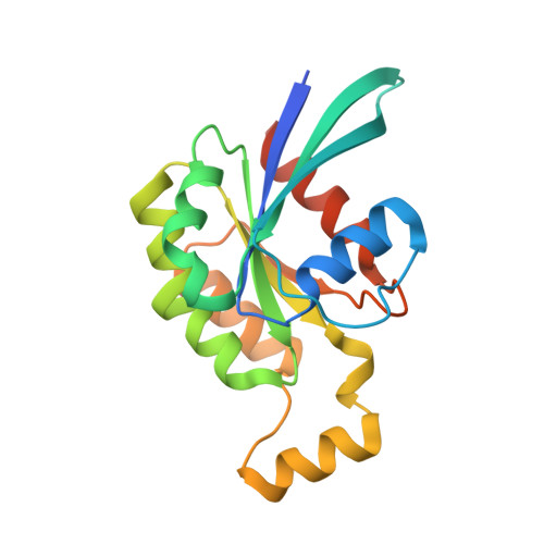
Cryo-EM structure of DOCK11 complexed with Cdc42(G15A) and Cdc42(Q61L)-GTP in the membrane-bound state


Experimental Data Snapshot

  • Method: ELECTRON MICROSCOPY
  • Resolution: 6.07 Å
  • Aggregation State: PARTICLE 
  • Reconstruction Method: SINGLE PARTICLE 

Starting Model: experimental
View more details

wwPDB Validation 3D Report Full Report

Validation slider image for 22VG

This is version 1.0 of the entry. See complete history. 

Literature

Structural basis for membrane-dependent positive feedback regulation of DOCK11 by activated Cdc42.

Hisano, T., Shinoda, T., Kukimoto-Niino, M., Katsura, K., Kondo, R., Ishizuka-Katsura, Y., Miyata-Yabuki, Y., Hanada, K., Nakagawa, R., Nishikimi, A., Shirouzu, M.

(2026) Proc Natl Acad Sci U S A 123: e2607665123-e2607665123

  • DOI: https://doi.org/10.1073/pnas.2607665123
  • Primary Citation Related Structures: 
    22VD, 22VE, 22VF, 22VG

  • PubMed Abstract: 

    The guanine nucleotide exchange factor DOCK11 is widely expressed across tissues and activates Cdc42, a Rho family GTPase that regulates cytoskeletal remodeling and cell polarity. Loss-of-function mutations in DOCK11 have recently been linked to inborn errors of immunity. DOCK11 has also been implicated in facilitating hepatitis B virus infection in hepatocytes and has emerged as a potential target for therapeutic interventions. A positive feedback mechanism has been proposed for DOCK11-mediated Cdc42 activation; however, the structural basis for this regulation has remained unclear. Here we report the cryoelectron microscopy structures of full-length DOCK11 as an apo dimer, a binary complex with nucleotide-free Cdc42, and a ternary complex containing both nucleotide-free and activated Cdc42. DOCK11 predominantly adopts a U-shaped dimer that binds to nucleotide-free Cdc42 via the catalytic domain, whereas activated Cdc42 engages a distal site within the armadillo-repeat domain. Biochemical and cellular analyses demonstrate that binding of activated Cdc42 is critical for enhancing the membrane-dependent nucleotide exchange activity of DOCK11, promoting its localization to the plasma membrane, and facilitating intracellular activation of Cdc42. We also determined the structure of the membrane-bound ternary complex using membrane-coated grids. Strikingly, the ternary complex adopts an extended conformation that aligns multiple membrane-contact elements, revealing how activated Cdc42 stabilizes DOCK11 membrane binding for efficient exchange activity. Collectively, these findings provide a structural framework for the coordinated mechanism underlying membrane-dependent positive feedback in Rho GTPase signaling.


  • Organizational Affiliation: 
    • Laboratory for Protein Functional and Structural Biology, RIKEN Center for Integrative Medical Sciences, Yokohama 230-0045, Kanagawa, Japan.

Macromolecule Content 

  • Total Structure Weight: 563.73 kDa 
  • Atom Count: 33,818 
  • Modeled Residue Count: 4,194 
  • Deposited Residue Count: 4,938 
  • Unique protein chains: 3

Macromolecules

Find similar proteins by:|  3D Structure
Entity ID: 1
MoleculeChains  Sequence LengthOrganismDetailsImage
Dedicator of cytokinesis protein 11A,
D [auth B]
2,079Homo sapiensMutation(s): 0 
Gene Names: DOCK11, ZIZ2
UniProt & NIH Common Fund Data Resources
Find proteins for Q5JSL3 (Homo sapiens)
Explore Q5JSL3 
Go to UniProtKB:  Q5JSL3
PHAROS:  Q5JSL3
GTEx:  ENSG00000147251 
Entity Groups
Sequence Clusters30% Identity50% Identity70% Identity90% Identity95% Identity100% Identity
UniProt GroupQ5JSL3
Sequence Annotations
Expand
Reference Sequence
Find similar proteins by:|  3D Structure
Entity ID: 2
MoleculeChains  Sequence LengthOrganismDetailsImage
Cell division control protein 42 homologB [auth C],
E [auth D]
195Homo sapiensMutation(s): 1 
Gene Names: CDC42
EC: 3.6.5.2
UniProt & NIH Common Fund Data Resources
Find proteins for P60953 (Homo sapiens)
Explore P60953 
Go to UniProtKB:  P60953
PHAROS:  P60953
GTEx:  ENSG00000070831 
Entity Groups
Sequence Clusters30% Identity50% Identity70% Identity90% Identity95% Identity100% Identity
UniProt GroupP60953
Sequence Annotations
Expand
Reference Sequence
Find similar proteins by:|  3D Structure
Entity ID: 3
MoleculeChains  Sequence LengthOrganismDetailsImage
Cell division control protein 42 homologC [auth E],
F
195Homo sapiensMutation(s): 1 
Gene Names: CDC42
EC: 3.6.5.2
UniProt & NIH Common Fund Data Resources
Find proteins for P60953 (Homo sapiens)
Explore P60953 
Go to UniProtKB:  P60953
PHAROS:  P60953
GTEx:  ENSG00000070831 
Entity Groups
Sequence Clusters30% Identity50% Identity70% Identity90% Identity95% Identity100% Identity
UniProt GroupP60953
Sequence Annotations
Expand
Reference Sequence

Experimental Data & Validation

Experimental Data

  • Method: ELECTRON MICROSCOPY
  • Resolution: 6.07 Å
  • Aggregation State: PARTICLE 
  • Reconstruction Method: SINGLE PARTICLE 
EM Software:
TaskSoftware PackageVersion
RECONSTRUCTIONcryoSPARC
MODEL REFINEMENTPHENIX1.21_5207

Structure Validation

View Full Validation Report


Entry History 

& Funding Information

Deposition Data


Funding OrganizationLocationGrant Number
Japan Science and TechnologyJapanJPMJCR22E3
Other governmentNational Center for Geriatrics and Gerontology, 24-7
Other privateBiology of Intracellular Environments

Revision History  (Full details and data files)

  • Version 1.0: 2026-10-21
    Type: Initial release