Download Mendeley
Perspectives for pharmaceutical screening at XFEL sources using the example of Lassa virus endonuclease.
Falke, S., Reinke, P.Y.A., Rosenberg, D., Fischer, P., Meyer, J., Galchenkova, M., Tolstikova, A., Mariani, V., Senst, J.M., Lewe, P., Witt, S., Wagner, A., Chapman, H.N., Hunter, M., Gunther, S., Meents, A.(2026) Acta Crystallogr D Struct Biol
- PubMed: 42789322 Search on PubMed
- DOI: https://doi.org/10.1107/S2059798326009435
- Primary Citation Related Structures:
28JG, 28JH, 28JI, 28UA, 28UB, 28UC, 29IK, 29IL, 29IM - PubMed Abstract:
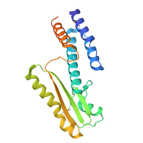
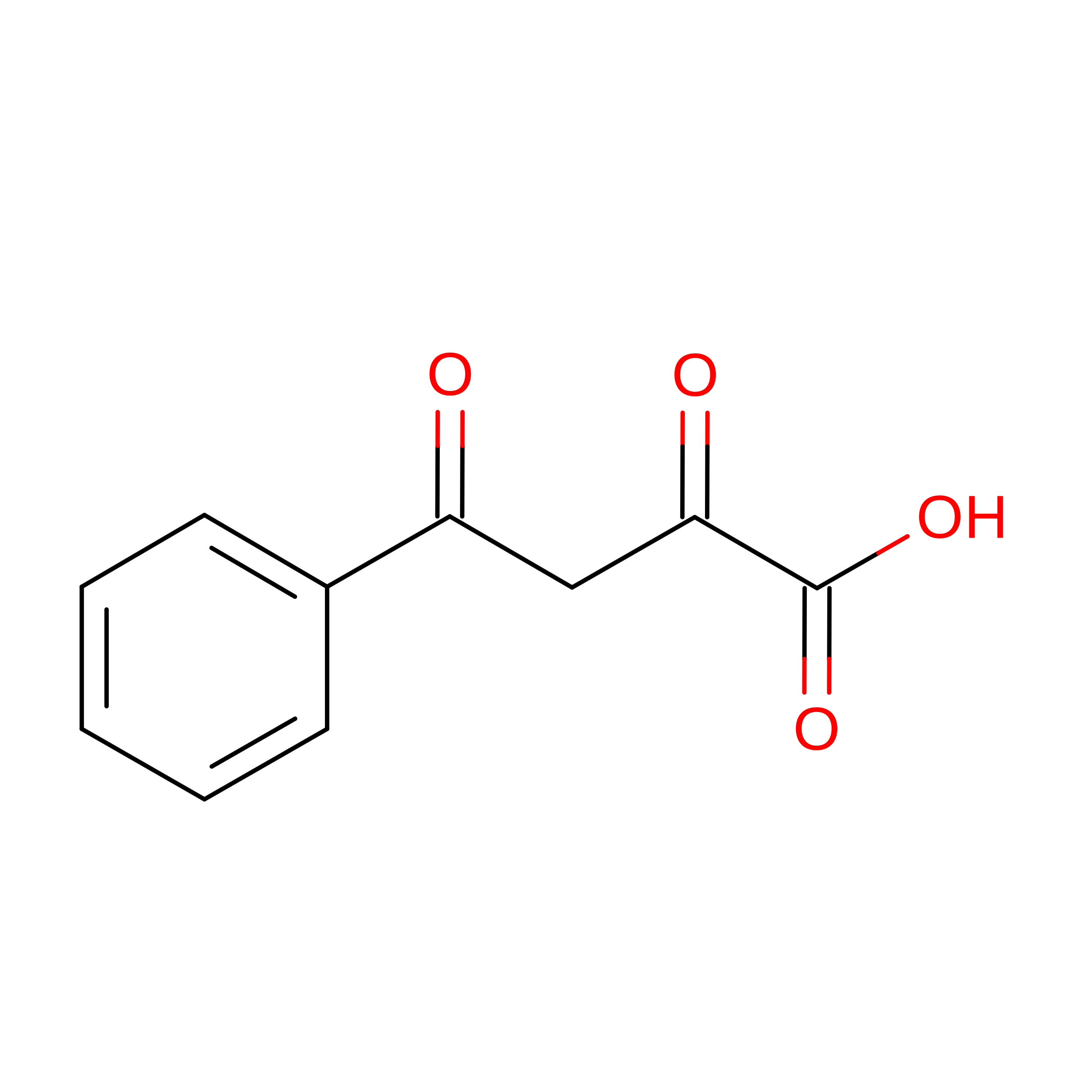
Emerging human pathogenic RNA viruses such as Lassa virus (LASV) and closely related viruses such as Hantavirus and Andes virus continue to be a major health threat globally. Treatment options for infections are severely limited. The widely conserved cap-snatching endonuclease of LASV (LASVendoN) is therefore used as an interesting drug target for our X-ray compound-screening experiments. Considering recent improvements in instrumentation and serial crystallography method development, different approaches may be used to facilitate pharmaceutical compound screening. Current-generation X-ray free-electron laser (XFEL) sources hold promise for obtaining higher quality diffraction data and achieving a high sample throughput using fixed-target serial femtosecond X-ray crystallography. We collected serial fixed-target X-ray diffraction data at Linac Coherent Light Source and at the synchrotron source PETRA III for comparison and solved the crystal structures of 2,4-dioxo-4-phenylbutanoic acid (DPBA) and baloxavir acid (BXA) binding to the active site of LASVendoN. These solved structures represent a reasonable starting point for drug development. In addition to room-temperature diffraction data, conventional single-crystal diffraction data were recorded at 100 K for comparison. To analyse the sample throughput in a compound-screening experiment at an XFEL, we further present data for the bacterial fosfomycin-resistance protein A with six ligands previously identified in a synchrotron screening experiment. In summary, the XFEL and synchrotron data-collection approaches have proven to be useful for compound screening with minor differences in achievable data quality and resolution. The individual experimental setup, detector and potentially the use of beam sweeping at XFELs at close-to-physiological temperatures combined with reasonable sample consumption and minimized radiation damage are considered to be advantageous for high-throughput drug development.
- Center for Free-Electron Laser Science CFEL, Deutsches Elektronen-Synchrotron DESY, Notkestrasse 85, 22607 Hamburg, Germany.
Organizational Affiliation: